ตำรามาตรฐานยาสมุนไพรไทย
Thai Herbal Pharmacopoeia
สำนักยาและวัตถุเสพติด กรมวิทยาศาสตร์การแพทย์ กระทรวงสาธารณสุข
Bureau of Drug and Narcotic, Department of Medical Sciences, Ministry of Public Healthสำนักยาและวัตถุเสพติด กรมวิทยาศาสตร์การแพทย์ กระทรวงสาธารณสุข
Bureau of Drug and Narcotic, Department of Medical Sciences, Ministry of Public Health(Tinospora crispa (L.) Hook.f. & Thomson)
(Nelumbo nucifera Gaertn.)
(Centella asiatica (L.) Urb.)
(Centella Dry Extract)
(Centella Cream)
(Mesua ferrea L.)
(Piper sarmentosum Roxb.)
(Piper sarmentosum Roxb.)
(Pterocarpus santalinus L. f.)
(Santalum album L.)
(Senna tora (L.) Roxb.)
(Senna alata (L.) Roxb.)
(Senna Alata Tea)
(Piper retrofractum Vahl)
(Myristica fragrans Houtt)
(Andrographis paniculata (Burm. f.) Nees)
(Andrographis Capsules)
(Allium ascalonicum L.)
(Ocimum tenuiflorum L.)
(Curcuma longa L.)
(Turmeric Capsules)
(Turmeric Dry Extract)
(Turmeric Dry Extract Capsules)
(Arcangelisia flava (L.) Merr.)
(Curcuma sp.)
Harrisonia perforata (Blanco) Merr.
(Aristolochia pierrei Lecomte)
(Zingiber officinale Roscoe)
(Ginger Capsules)
(Ginger Tea)
(Cassia fistula L.)
(Nardostachys jatamansi (D. Don) DC.)
(Angelica sinensis (Oliv.) Diels)
Artemisia annua L.
(Ligusticum sinense Oliv. cv. Chuanxiong)
(Neopicrorhiza scrophulariiflora Pennell)
(Atractylodes lancea (Thunb.) DC.)
(Aucklandia lappa Decne)
(Terminalia chebula Retz.)
(Angelica dahurica (Hoffm.) Benth. & Hook. f. ex Franch. & Sav. var. dahurica)
(Kaempferia parviflora Wall. ex Baker)
(Hibiscus sabdariffa L.)
(Roselle Tea)
(Allium sativum L.)
(Zingiber zerumbet (L.) Sm.)
(Wurfbainia testacea (Ridl.) Škorničk.& A. D. Poulsen)
(Cannabis sativa L.)
(Myristica fragrans Houtt)
(Dracaena cochinchinensis (Lour.) S. C. Chen)
(Ficus racemosa L.)
(Hyptis suaveolens (L.) Poit.)
Clerodendrum indicum (L.) Kuntze
(Phyllanthus emblica L.)
(Citrus hystrix DC.)
(Citrus hystrix DC.)
(Areca catechu L.)
(Momordica charantia L.)
Moringa oleifera Lam.
(Aegle marmelos (L.) Corrêa)
(Solanum trilobatum L.)
(Morus alba L.)
Gynostemma pentaphyllum(Thunb.)
Makino
(Clinacanthus nutans (Burm. f.) Lindau)
(Cissus quadrangularis L.)
(Mimusops elengi L.)
(Zingiber montanum (J. König) Link. ex A. Dietr.)
(Piper betle L.)
(Capsicum annuum L.)
(Capsicum Oleoresin)
(Capsicum Gel)
(Piper nigrum L.)
(Piper nigrum L.)
(Eurycoma longifolia Jack)
(Thunbergia laurifolia Lindl.)
(Piper wallichii (Miq.) Hand.-Mazz.)
Senna garrettiana (Craib) H. S. Irwin & Barneby
(Terminalia bellirica (Gaertn.) Roxb.)
(Terminalia chebula Retz.)
(Caesalpinia bonduc (L.) H. Roxb.)
(Tarlmounia elliptica (DC.) H. Rob., S. C. Keeley, Skvaria & R. Chan)
(Hog Creeper Vine Dry Extract Capsiles)
(Hog Creeper Vine Dry Extract)
(Brachypterum scandens (Roxb.) Miq.)
(Lepidium sativum L.)
(Nigella sativa L.)
(Cuminum cyminum L.)
(Foeniculum vulgare Mill.)
(Plantago ovata Forssk.)
(Pimpinella anisum L.)
(Carum carvi L.)
(Anethum graveolens L.)
(Trachyspermum ammi (L.) Sprague)
Albizia procera (Roxb.) Benth.
(Acorus calamus L.)
(Tiliacora triandra (Colebr.) Diels)
Cyanthillium cinereum (L.) H. Rob.
(Orthosiphon aristatus (Blume) Miq.)
Murdannia loriformis (Hassk.) R. S. Rao & Kammathy
(Capparis micracantha DC.)
(Chrysopogon zizanioides (L.) Roberty)
(Cyperus rotundus L.)
(Cannabis sativa L.)
(Syzygium aromaticum (L.) Merr. & L. M. Perry)
(Boesenbergia rotunda (L.) Mansf.)
(Acanthus ebracteatus Vahl)
(Acanthus ilicifolius L.)
(Kaempferia galanga L.)
(Curcuma comosa Roxb.)
Betula alnoides Buch.-Ham. ex D. Don
Cannabis sativa L.
Carthamus tinctorius L
Mitragyna speciosa (Korth.) Havil
Mallotus repandus (Rottler) Müll. Arg
Azadirachta indica A. Juss. var. siamensis Valeton
Azadirachta indica A. Juss. var. siamensis Valeton
Punica granatum L.
Rhinacanthus nasutus (L.) Kurz
Baliospermum solanifolium (Burm.) Suresh
Curcuma aeruginosa Roxb
Boesenbergia kingii Mood & L. M. Prince
Senegalia rugata (Lam.) Britton & Rose
Acacia concinna (Willd.) DC.
Senegalia rugata (Lam.) Britton & Rose
Acacia concinna (Willd.) DC.
Senna alexandriana Mill. var. alexandriana
Cassia acutifolia Delile, Cassia angustifolia Vahl
Butea superba Roxb. ex Willd.
[Plaso superba (Roxb. ex Willd.) Kuntze, Rudolphia superba (Roxb. ex Willd.) Poir.
Pueraria candollei Graham
ex Benth. var. mirifica (Airy Shaw & Suvat.) Niyomdham
Streblus asper Lour.
Suregada multiflora (A. Juss.) Baill. (Gelonium
multiflorum A. Juss.
Plumbago zeylanica L.
Plumbago indica L.
Biancaea sappan (L.) Tod.
Ziziphus attopensis Pierre
Streblus asper Lour.
Justicia gendarussa Burm. f.
Enhalus acoroides (L. f.) Royle
Bridelia ovata Decne.
Tamarindus indica L.
Citrus × aurantiifolia (Christm.) Swingle
Garcinia mangostana L.
Blumea balsamifera (L.) DC
Persicaria odorata (Lour.) Soják
Zingiber montanum (J. König) Link ex A. Dietr.
Mammea siamensis (Miq.) T. Anderson
Citrus maxima (Burm.) Merr.
Citrus × aurantium L. ‘Som Sa'
Punica granatum L.
Rhinacanthus nasutus (L.) Kurz
Forest Siris Bark is the dried stem bark of Albizia procera (Roxb.) Benth. [Acacia procera (Roxb.) Willd, Mimosa procera Roxb.] (Family Leguminosae-Mimosoideae), Herbarium Specimen Number: DMSC 5251, Crude Drug Number: DMSc 1177.
Constituents Forest Siris Bark contains phenolics (e.g., (+)-catechin, protocatechuic acid). It also contains flavonoids (e.g., quercetin, rutin), triterpenoids, saponins, tannins, etc.
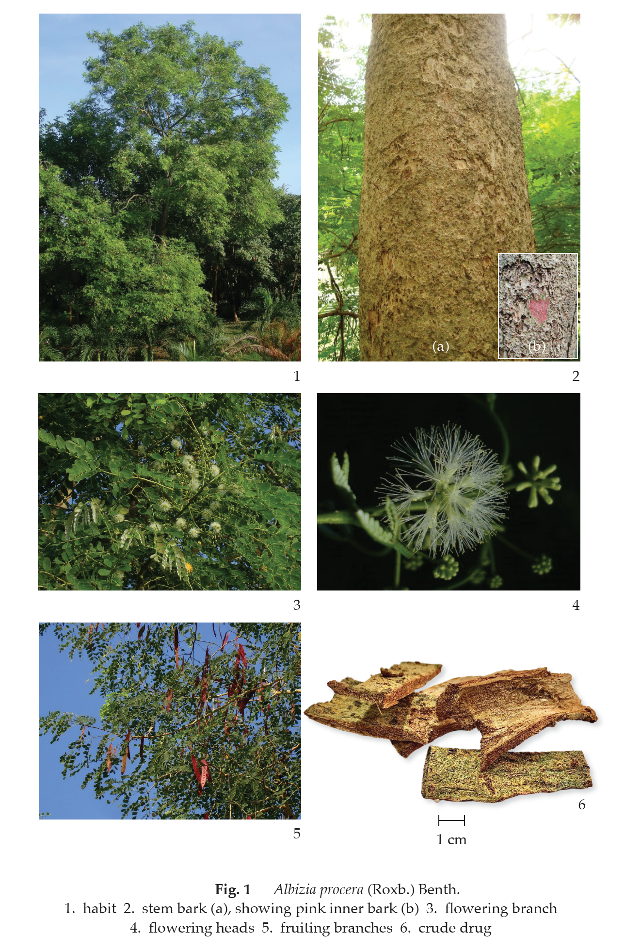
Description of the plant (Fig. 1) Tree up to 15 m tall; bark pale greyish green, yellowish brown to brown, rough; inner bark pink; branchlet terete, glabrous. Leaves bipinnate; rachis 10 to 45 cm long, with narrowly elliptic gland 0.6 to 1 cm long at base; pinnae 2 to 5 pairs, 12 to 20 cm long; leaflets (4‒)5 to 11(‒16) pairs, opposite; petiolule very short; blade ovate to subrhomboid, 3 to 4.5 cm long, 1.2 to 2.2 cm wide, apex obtuse, rounded, emarginate with a tiny mucro, base asymmetrical, truncate or cuneate, chartaceous, faintly puberulous on both surfaces. Inflorescence panicle, 15 to 30 peduncled heads, axillary or terminal, up to 30 cm long; peduncle 1.5 to 2.5 cm long. Flower sessile, uniform; calyx 2 to 3 mm long, tubular to narrowly funnel-shaped, glabrous, lobes 5-toothed, 0.7 to 1.2 mm long; corolla whitish, funnel-shaped, 6 to 6.5 mm long, tube glabrous, lobes 5, elliptic, 2 to 2.5 mm long, acute and puberulous at apex; stamens numerous; lower part of filament united into staminal tube, longer than corolla tube; ovary superior, subsessile, labrous. Fruit dehiscent pod, flat, 10 to 20 cm long, 1.5 to 2.5 cm wide, shortly stalked, reddish brown to brown, glabrous, with distinct marks over the seed. Seeds 6 to 12, elliptic to obovoid, about 7.5 mm long, about 4.5 mm wide, flat.
Description Odour, mild; taste, slightly bitter.
Macroscopical (Fig. 1) Bark, irregularly shaped; outer part rough, yellowish brown to brown; middle part, pinkish; inner part smooth, yellowish to straw-coloured.
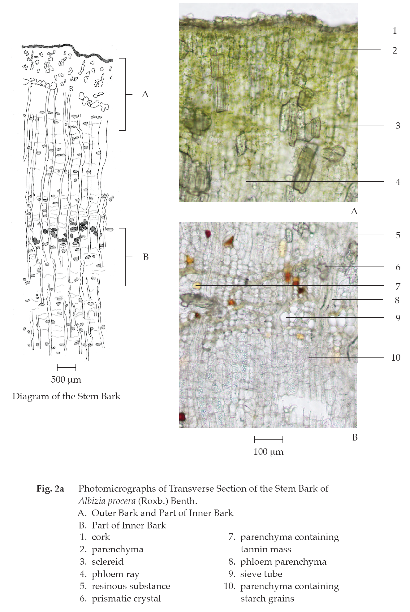
Microscopical (Figs. 2a, 2b) Transverse section of the stem bark shows outer and inner barks. Outer bark: layers of yellow thin-walled rectangular cork cells; groups of lignified sclereids, some containing tannin mass and resinous substance; and parenchyma, some containing tannin mass and resinous substance. Inner bark: phloem parenchyma, some containing starch grains, prismatic crystals, oil droplets, resinous substance, and tannin mass; sieve tube; phloem rays, some containing starch grains; and phloem fibres.
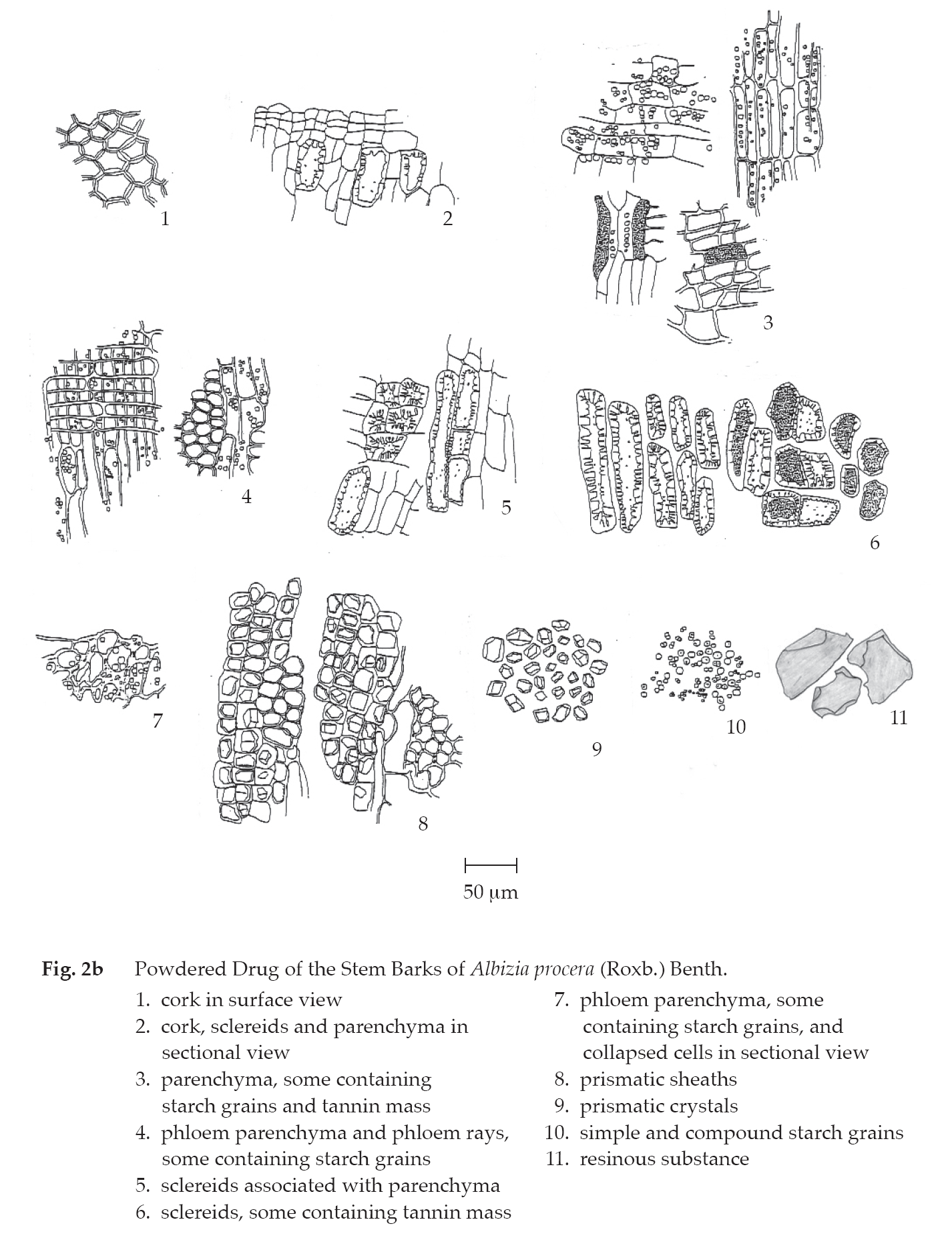
Forest Siris Bark in powder possesses the diagnostic microscopical characters of the unground drug. Prismatic sheath and various shapes and sizes of sclereids, some containing tannin mass and resinous substance, are frequently observed.
Packaging and storage Forest Siris Bark shall be kept in well-closed containers, protected from light, and stored in a dry place.
Identification
A. Reflux 500 mg of the sample, in fine powder, with 10 mL of methanol for 15 minutes and filter. To 2 mL of the filtrate, add 1 or 2 pieces of magnesium ribbon, mix with 4 or 5 drops of hydrochloric acid and warm on a water-bath for 5 minutes: a red colour develops.
B. Heat 500 mg of the sample, in fine powder, with 10 mL of water on a water-bath for 15 minutes and filter. Shake 5 mL of the filtrate in a screw-capped tube for 15 seconds: a persisting foam is produced for over 30 minutes.
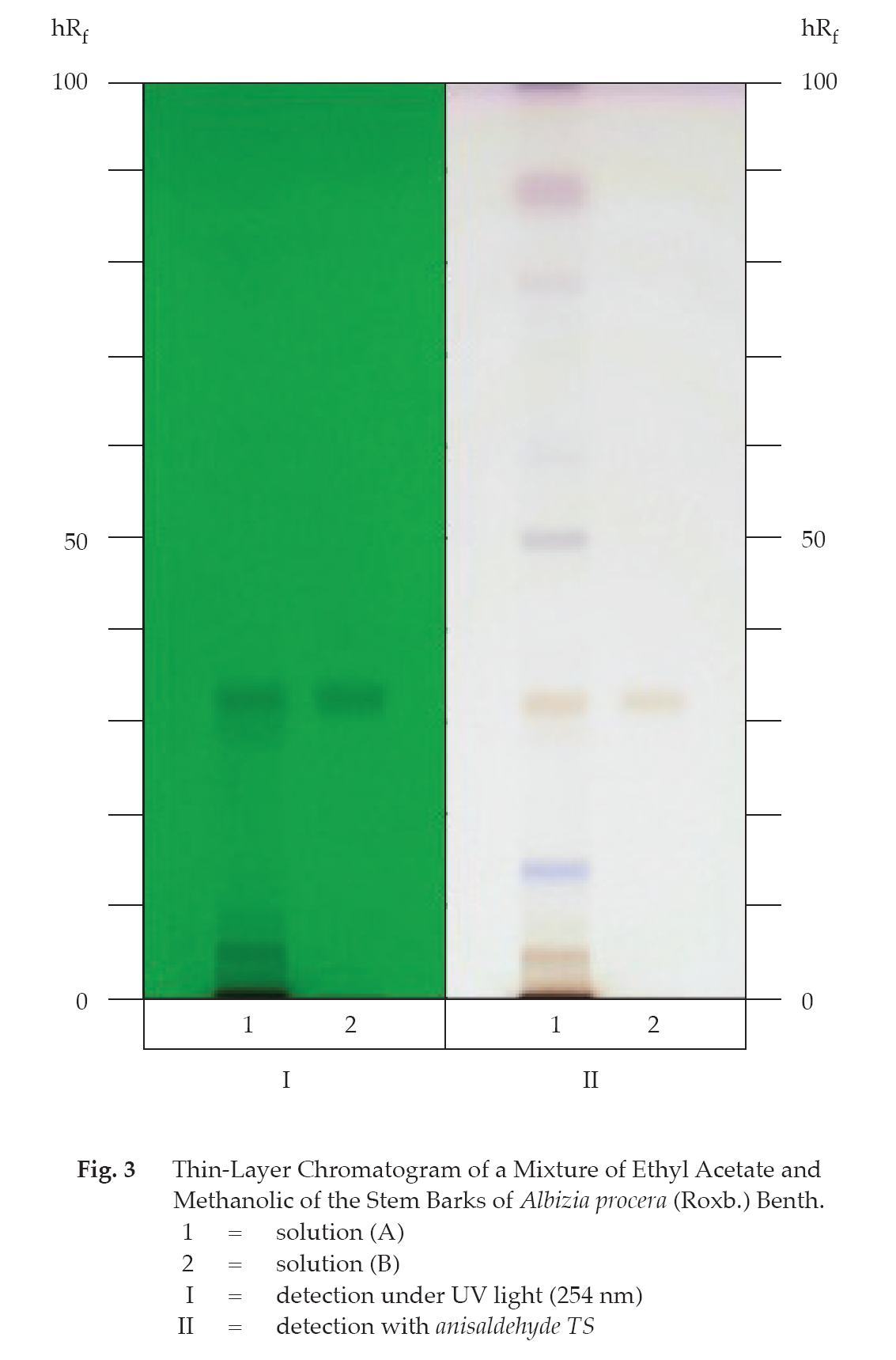
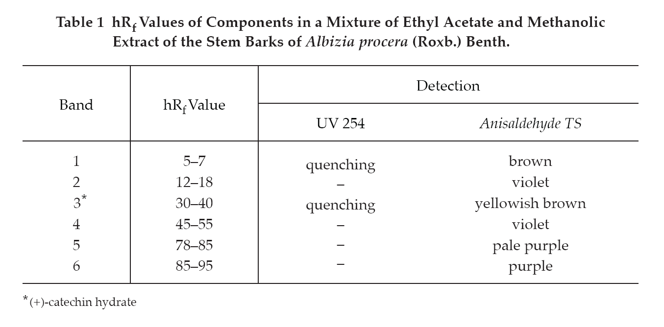
C. Carry out the test as described in the “Thin-Layer Chromatography” (Appendix 3.1), using silica gel F254 as the coating substance and a mixture of 50 volumes of toluene, 50 volumes of ethyl acetate and 5 volumes of formic acid as the mobile phase and allowing the solvent front to ascend 10 cm above the line of application. Apply separately to the plate as bands of 8 mm, 5 µL of solution (A) and 1 µL of solution (B). Prepare solution (A) by refluxing 500 mg of the sample, in fine powder, with 10 mL of a mixture of equal volumes of ethyl acetate and methanol for 10 minutes and filtering. Evaporate the filtrate to dryness and dissolve the residue in 2 mL of methanol. For solution (B), dissolve 1 mg of (+)-catechin hydrate in 1 mL of methanol. After removal of the plate, allow it to dry in air and examine the plate under ultraviolet light (254 nm), marking the quenching bands. The chromatogram obtained from solution (A) shows a quenching band (hRf value 30 to 40) corresponding to the (+)-catechin hydrate band from solution (B); other quenching bands are also observed. Subsequently spray the plate with anisaldehyde TS and heat at 105° for 3 minutes; the band due to (+)-catechin hydrate is yellowish brown. Several other bands of different colours are observed. (Table 1); see also Fig. 3.
Loss on drying Not more than 10.0 per cent w/w after drying at 105° to constant weight (Appendix 4.15).
Foreign matter Not more than 2.0 per cent w/w (Appendix 7.2).
Total ash Not more than 11.0 per cent w/w (Appendix 7.7).
Ethanol-soluble extractive Not less than 20.0 per cent w/w (Appendix 7.12).
Water-soluble extractive Not less than 18.0 per cent w/w (Appendix 7.12).