ตำรามาตรฐานยาสมุนไพรไทย
Thai Herbal Pharmacopoeia
สำนักยาและวัตถุเสพติด กรมวิทยาศาสตร์การแพทย์ กระทรวงสาธารณสุข
Bureau of Drug and Narcotic, Department of Medical Sciences, Ministry of Public Healthสำนักยาและวัตถุเสพติด กรมวิทยาศาสตร์การแพทย์ กระทรวงสาธารณสุข
Bureau of Drug and Narcotic, Department of Medical Sciences, Ministry of Public Health(Tinospora crispa (L.) Hook.f. & Thomson)
(Nelumbo nucifera Gaertn.)
(Centella asiatica (L.) Urb.)
(Centella Dry Extract)
(Centella Cream)
(Mesua ferrea L.)
(Piper sarmentosum Roxb.)
(Piper sarmentosum Roxb.)
(Pterocarpus santalinus L. f.)
(Santalum album L.)
(Senna tora (L.) Roxb.)
(Senna alata (L.) Roxb.)
(Senna Alata Tea)
(Piper retrofractum Vahl)
(Myristica fragrans Houtt)
(Andrographis paniculata (Burm. f.) Nees)
(Andrographis Capsules)
(Allium ascalonicum L.)
(Ocimum tenuiflorum L.)
(Curcuma longa L.)
(Turmeric Capsules)
(Turmeric Dry Extract)
(Turmeric Dry Extract Capsules)
(Arcangelisia flava (L.) Merr.)
(Curcuma sp.)
Harrisonia perforata (Blanco) Merr.
(Aristolochia pierrei Lecomte)
(Zingiber officinale Roscoe)
(Ginger Capsules)
(Ginger Tea)
(Cassia fistula L.)
(Nardostachys jatamansi (D. Don) DC.)
(Angelica sinensis (Oliv.) Diels)
Artemisia annua L.
(Ligusticum sinense Oliv. cv. Chuanxiong)
(Neopicrorhiza scrophulariiflora Pennell)
(Atractylodes lancea (Thunb.) DC.)
(Aucklandia lappa Decne)
(Terminalia chebula Retz.)
(Angelica dahurica (Hoffm.) Benth. & Hook. f. ex Franch. & Sav. var. dahurica)
(Kaempferia parviflora Wall. ex Baker)
(Hibiscus sabdariffa L.)
(Roselle Tea)
(Allium sativum L.)
(Zingiber zerumbet (L.) Sm.)
(Wurfbainia testacea (Ridl.) Škorničk.& A. D. Poulsen)
(Cannabis sativa L.)
(Myristica fragrans Houtt)
(Dracaena cochinchinensis (Lour.) S. C. Chen)
(Ficus racemosa L.)
(Hyptis suaveolens (L.) Poit.)
Clerodendrum indicum (L.) Kuntze
(Phyllanthus emblica L.)
(Citrus hystrix DC.)
(Citrus hystrix DC.)
(Areca catechu L.)
(Momordica charantia L.)
Moringa oleifera Lam.
(Aegle marmelos (L.) Corrêa)
(Solanum trilobatum L.)
(Morus alba L.)
Gynostemma pentaphyllum(Thunb.)
Makino
(Clinacanthus nutans (Burm. f.) Lindau)
(Cissus quadrangularis L.)
(Mimusops elengi L.)
(Zingiber montanum (J. König) Link. ex A. Dietr.)
(Piper betle L.)
(Capsicum annuum L.)
(Capsicum Oleoresin)
(Capsicum Gel)
(Piper nigrum L.)
(Piper nigrum L.)
(Eurycoma longifolia Jack)
(Thunbergia laurifolia Lindl.)
(Piper wallichii (Miq.) Hand.-Mazz.)
Senna garrettiana (Craib) H. S. Irwin & Barneby
(Terminalia bellirica (Gaertn.) Roxb.)
(Terminalia chebula Retz.)
(Caesalpinia bonduc (L.) H. Roxb.)
(Tarlmounia elliptica (DC.) H. Rob., S. C. Keeley, Skvaria & R. Chan)
(Hog Creeper Vine Dry Extract Capsiles)
(Hog Creeper Vine Dry Extract)
(Brachypterum scandens (Roxb.) Miq.)
(Lepidium sativum L.)
(Nigella sativa L.)
(Cuminum cyminum L.)
(Foeniculum vulgare Mill.)
(Plantago ovata Forssk.)
(Pimpinella anisum L.)
(Carum carvi L.)
(Anethum graveolens L.)
(Trachyspermum ammi (L.) Sprague)
Albizia procera (Roxb.) Benth.
(Acorus calamus L.)
(Tiliacora triandra (Colebr.) Diels)
Cyanthillium cinereum (L.) H. Rob.
(Orthosiphon aristatus (Blume) Miq.)
Murdannia loriformis (Hassk.) R. S. Rao & Kammathy
(Capparis micracantha DC.)
(Chrysopogon zizanioides (L.) Roberty)
(Cyperus rotundus L.)
(Cannabis sativa L.)
(Syzygium aromaticum (L.) Merr. & L. M. Perry)
(Boesenbergia rotunda (L.) Mansf.)
(Acanthus ebracteatus Vahl)
(Acanthus ilicifolius L.)
(Kaempferia galanga L.)
(Curcuma comosa Roxb.)
Betula alnoides Buch.-Ham. ex D. Don
Cannabis sativa L.
Carthamus tinctorius L
Mitragyna speciosa (Korth.) Havil
Mallotus repandus (Rottler) Müll. Arg
Azadirachta indica A. Juss. var. siamensis Valeton
Azadirachta indica A. Juss. var. siamensis Valeton
Punica granatum L.
Rhinacanthus nasutus (L.) Kurz
Baliospermum solanifolium (Burm.) Suresh
Curcuma aeruginosa Roxb
Boesenbergia kingii Mood & L. M. Prince
Senegalia rugata (Lam.) Britton & Rose
Acacia concinna (Willd.) DC.
Senegalia rugata (Lam.) Britton & Rose
Acacia concinna (Willd.) DC.
Senna alexandriana Mill. var. alexandriana
Cassia acutifolia Delile, Cassia angustifolia Vahl
Butea superba Roxb. ex Willd.
[Plaso superba (Roxb. ex Willd.) Kuntze, Rudolphia superba (Roxb. ex Willd.) Poir.
Pueraria candollei Graham
ex Benth. var. mirifica (Airy Shaw & Suvat.) Niyomdham
Streblus asper Lour.
Suregada multiflora (A. Juss.) Baill. (Gelonium
multiflorum A. Juss.
Plumbago zeylanica L.
Plumbago indica L.
Biancaea sappan (L.) Tod.
Ziziphus attopensis Pierre
Streblus asper Lour.
Justicia gendarussa Burm. f.
Enhalus acoroides (L. f.) Royle
Bridelia ovata Decne.
Tamarindus indica L.
Citrus × aurantiifolia (Christm.) Swingle
Garcinia mangostana L.
Blumea balsamifera (L.) DC
Persicaria odorata (Lour.) Soják
Zingiber montanum (J. König) Link ex A. Dietr.
Mammea siamensis (Miq.) T. Anderson
Citrus maxima (Burm.) Merr.
Citrus × aurantium L. ‘Som Sa'
Punica granatum L.
Rhinacanthus nasutus (L.) Kurz
Sweet Flag Root is the dried rhizome of Acorus calamus L. (Family Acoraceae), Herbarium Specimen Number: DMSC 890.
Constituents Sweet Flag Root contains volatile oil comprising β-asarone, cis-methyl isoeugenol, α-and r-asarone, asarylaldehyde, acorone,acoroxide, acorin, calamene, linalool, calamol, calameone, eugenol, methyl eugenol, azulene, pinene, cineole, camphor, and others.
Other constituents include sesquiterpenoids (e.g., acoragermacrone, acolamone and iso-acolamone), acoric acid, tannin, and resin.
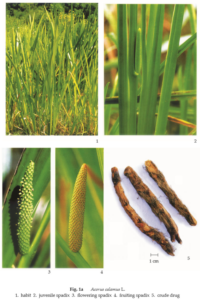
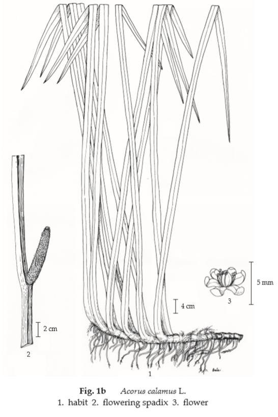
Description of the plant (Figs. 1a, 1b) Aquatic perennial herb with long creeping horizontal rhizome, growing in shallow water, marshes, or swamps. Leaves equitant, grass-like or sword-shaped, up to 2 m long, 1 to 3.5 cm wide, long and slender without distinction of petiole and finely parallel-veined. Inflorescence naked spike-like; spadix up to 10 cm long, projecting out and upwards from the leaf-like scape and spathe. Flower tiny, densely arranged, bisexual with perianth of 6 concave segments; stamens 6; ovary 2- to 4-celled with 2 to several ovules. Fruit baccate.
Description Odour, sweet and aromatic; taste, bitter and pungent.
Macroscopical (Fig. 1a) The rhizome is longitudinally splitted into subcylindrical pieces, 5 to 20 cm long, 0.5 to 1.5 cm in diameter. The unpeeled drug is covered in a thin brown cork and is much shrunken and deeply longitudinally wrinkled. It bears on the upper surface triangular leaf scars and hair-like fibres of fibrovascular tissue, and on the under surface many small root scars. The peeled drug is cream-yellow in colour and exhibits fewer root scars. The rhizome breaks with a sharp fracture producing a granular, white, spongy surface internally. The section has a large stele separated by a yellowish line from the cortex. Both the stele and cortex are covered with small oval fibrovascular bundles.
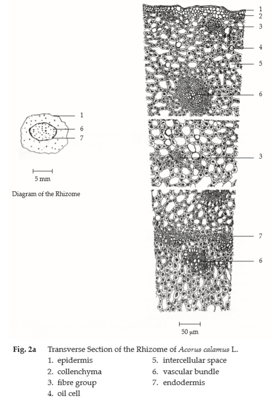
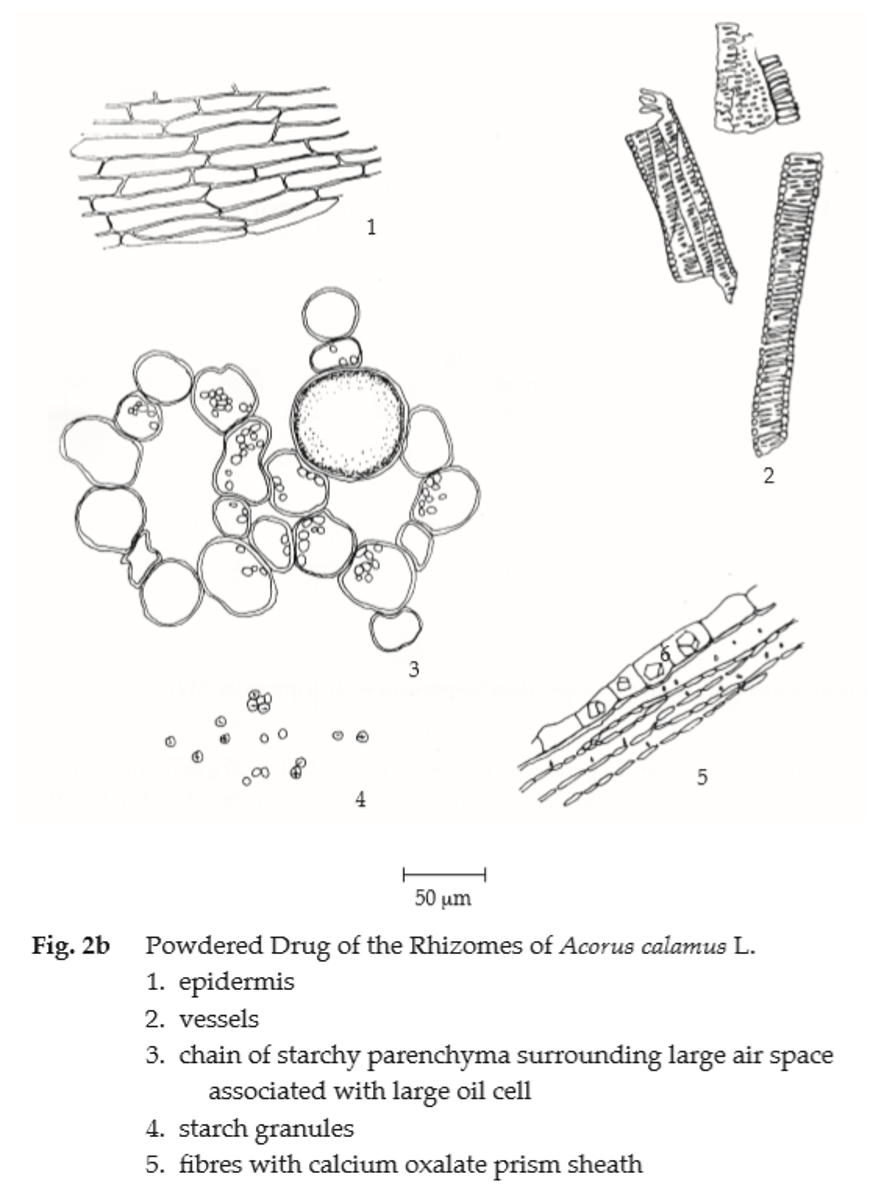
Microscopical (Figs. 2a, 2b) Transverse section of rhizome shows epidermis, a single layer of small thin-walled cells; outer region of the cortex, several layers of collenchyma; inner region of the cortex, chain of starchy parenchyma surrounding large air spaces with oil cells in each chain, fibrovascular bundles scattered throughout, surrounded with calcium oxalate prism sheath. Endodermis, a single layer of thin-walled elliptical cells. Stele, chain of starchy parenchyma surrounding large air spaces with yellowish brown oil cells, vascular bundles without fibre, scattered throughout, more numerously beneath the endodermis. Sweet Flag Root in powder possesses the diagnostic microscopical characters of the unground drug.
Warning For reasons of optimal drug safety, the use of β-asarone-rich races shall be avoided.
Additional information It is commonly used with other herbal drugs in Thai traditional herbal preparations.
Packaging and storage Sweet Flag Root shall be kept in well-closed containers, protected from light, and stored in a cool and dry place.
Identification
A. Reflux 1 g of the sample, in powder, with 25 mL of ethanol for 10 minutes and filter (solution 1). To 2 mL of solution 1, add a few drops of ninhydrin TS and heat in a water-bath: a violet colour is produced.
B. To 2 mL of solution 1, add a few drops of basic lead acetate TS: a white precipitate is produced.
C. Evaporate 2 mL of solution 1 to dryness, dissolve the residue in 2 mL of acetic anhydride, and add carefully 1 mL of sulfuric acid to form a layer: a brown ring forms at the zone of contact; a yellow colour develops in the upper layer and then gradually changes to dirty green.
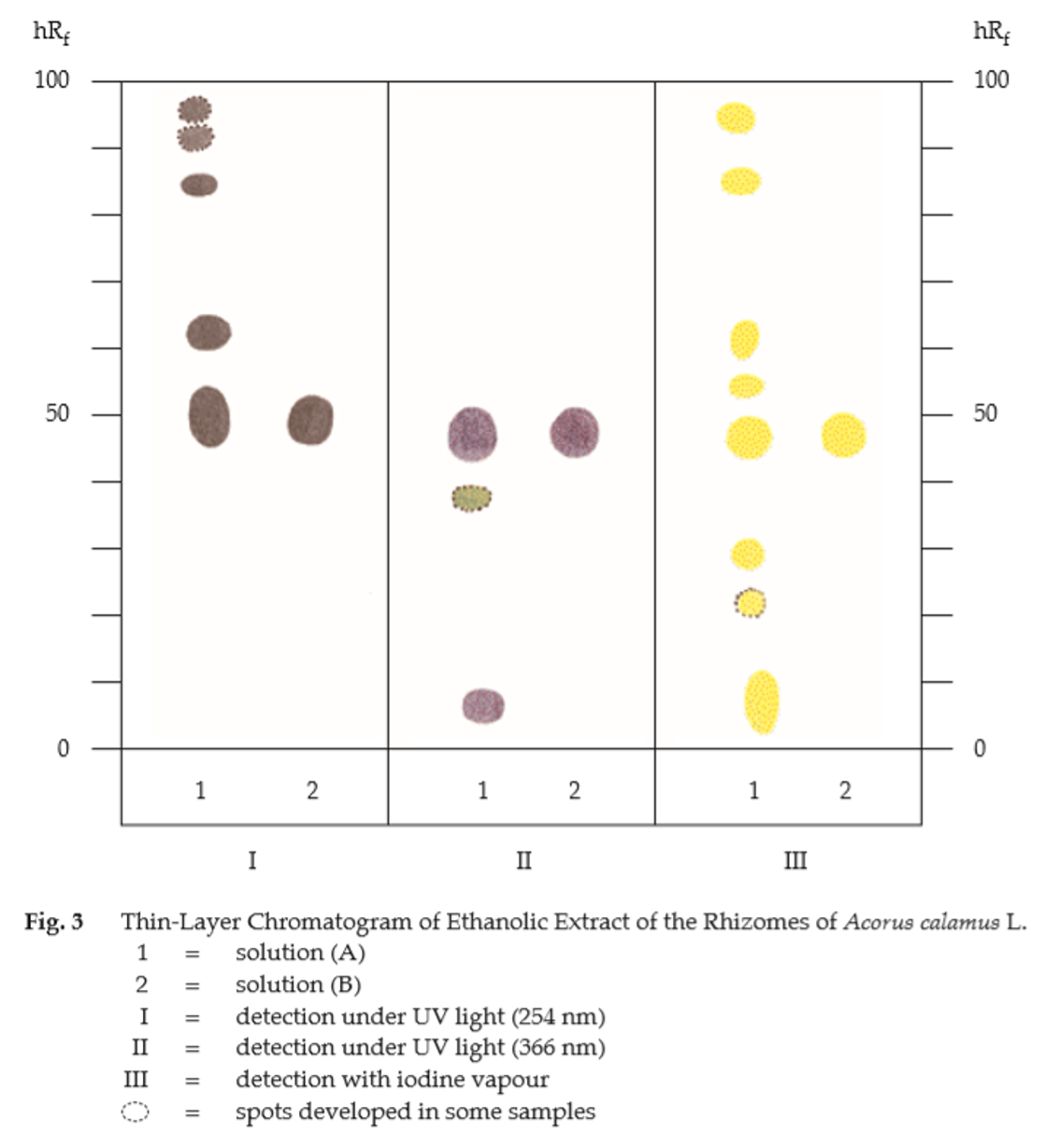
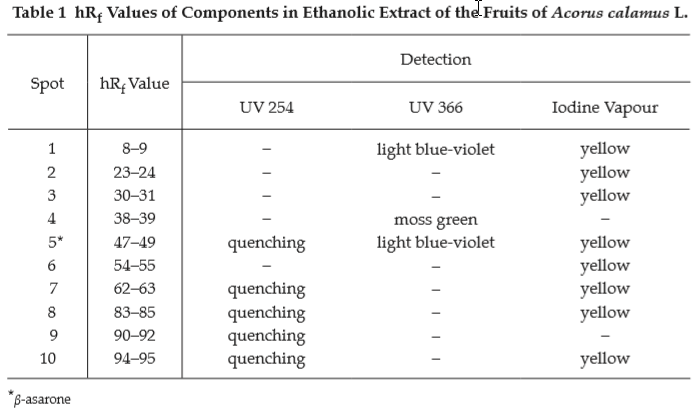
D. Carry out the test as described in the “Thin-Layer Chromatography” (Appendix 3.1), using silica gel GF254 as the coating substance and a mixture of 85 volumes of petroleum ether (boiling range, 40° to 60°) and 15 volumes of ethyl acetate as the mobile phase. Apply separately to the plate, 5 μL each of the following solutions. Prepare solution (A) by refluxing 1 g of the sample, in powder, with 25 mL of ethanol for 10 minutes, filtering, and evaporating to a volume of 2 mL. For solution (B), dissolve 2 mg of β-asarone in 1 mL of ethanol. After removal of the plate, allow it to dry in air and examine under ultraviolet light (254 nm), marking the quenching spots. The chromatogram obtained from solution (A) shows a quenching spot (hRf value 47 to 49) corresponding to the β-asarone spot from solution (B), and several spots of higher hRf values (Table 1); see also Fig. 3. Subsequently examine the plate under ultraviolet light (366 nm); the spot due to β-asarone shows a light blue-violet fluorescence. Other two spots of different colours are also observed (Table 1); see also Fig. 3. Expose the plate to iodine vapour; the spot corresponding to β-asarone is yellow. Other several spots are also observed (Table 1); see also Fig. 3.
Water Not more than 12.0 per cent v/w (Azeotropic Distillation Method, Appendix 4.12).
Foreign matter Not more than 2.0 per cent w/ w (Appendix 7.2).
Acid-insoluble ash Not more than 1.0 per cent w/w (Appendix 7.6).
Total ash Not more than 7.0 per cent w/w (Appendix 7.7).
Ethanol-soluble extractive Not less than 5.0 per cent w/w (Appendix 7.12).
Water-soluble extractive Not less than 11.0 per cent w/w (Appendix 7.12).
Volatile oil Not less than 1.2 per cent v/w (Appendix 7.3H). Use 20 g, in coarse powder, freshly prepared and accurately weighed. Use 150 mL of water as the distillation liquid and a 500 mL round-bottomed flask. Distil at a rate of 2 to 3 mL per minute for 5 hours. Use 2.0 mL of xylene in the graduated tube.
Dose 1 to 3 g.