ตำรามาตรฐานยาสมุนไพรไทย
Thai Herbal Pharmacopoeia
สำนักยาและวัตถุเสพติด กรมวิทยาศาสตร์การแพทย์ กระทรวงสาธารณสุข
Bureau of Drug and Narcotic, Department of Medical Sciences, Ministry of Public Healthสำนักยาและวัตถุเสพติด กรมวิทยาศาสตร์การแพทย์ กระทรวงสาธารณสุข
Bureau of Drug and Narcotic, Department of Medical Sciences, Ministry of Public Health(Tinospora crispa (L.) Hook.f. & Thomson)
(Nelumbo nucifera Gaertn.)
(Centella asiatica (L.) Urb.)
(Centella Dry Extract)
(Centella Cream)
(Mesua ferrea L.)
(Piper sarmentosum Roxb.)
(Piper sarmentosum Roxb.)
(Pterocarpus santalinus L. f.)
(Santalum album L.)
(Senna tora (L.) Roxb.)
(Senna alata (L.) Roxb.)
(Senna Alata Tea)
(Piper retrofractum Vahl)
(Myristica fragrans Houtt)
(Andrographis paniculata (Burm. f.) Nees)
(Andrographis Capsules)
(Allium ascalonicum L.)
(Ocimum tenuiflorum L.)
(Curcuma longa L.)
(Turmeric Capsules)
(Turmeric Dry Extract)
(Turmeric Dry Extract Capsules)
(Arcangelisia flava (L.) Merr.)
(Curcuma sp.)
Harrisonia perforata (Blanco) Merr.
(Aristolochia pierrei Lecomte)
(Zingiber officinale Roscoe)
(Ginger Capsules)
(Ginger Tea)
(Cassia fistula L.)
(Nardostachys jatamansi (D. Don) DC.)
(Angelica sinensis (Oliv.) Diels)
Artemisia annua L.
(Ligusticum sinense Oliv. cv. Chuanxiong)
(Neopicrorhiza scrophulariiflora Pennell)
(Atractylodes lancea (Thunb.) DC.)
(Aucklandia lappa Decne)
(Terminalia chebula Retz.)
(Angelica dahurica (Hoffm.) Benth. & Hook. f. ex Franch. & Sav. var. dahurica)
(Kaempferia parviflora Wall. ex Baker)
(Hibiscus sabdariffa L.)
(Roselle Tea)
(Allium sativum L.)
(Zingiber zerumbet (L.) Sm.)
(Wurfbainia testacea (Ridl.) Škorničk.& A. D. Poulsen)
(Cannabis sativa L.)
(Myristica fragrans Houtt)
(Dracaena cochinchinensis (Lour.) S. C. Chen)
(Ficus racemosa L.)
(Hyptis suaveolens (L.) Poit.)
Clerodendrum indicum (L.) Kuntze
(Phyllanthus emblica L.)
(Citrus hystrix DC.)
(Citrus hystrix DC.)
(Areca catechu L.)
(Momordica charantia L.)
Moringa oleifera Lam.
(Aegle marmelos (L.) Corrêa)
(Solanum trilobatum L.)
(Morus alba L.)
Gynostemma pentaphyllum(Thunb.)
Makino
(Clinacanthus nutans (Burm. f.) Lindau)
(Cissus quadrangularis L.)
(Mimusops elengi L.)
(Zingiber montanum (J. König) Link. ex A. Dietr.)
(Piper betle L.)
(Capsicum annuum L.)
(Capsicum Oleoresin)
(Capsicum Gel)
(Piper nigrum L.)
(Piper nigrum L.)
(Eurycoma longifolia Jack)
(Thunbergia laurifolia Lindl.)
(Piper wallichii (Miq.) Hand.-Mazz.)
Senna garrettiana (Craib) H. S. Irwin & Barneby
(Terminalia bellirica (Gaertn.) Roxb.)
(Terminalia chebula Retz.)
(Caesalpinia bonduc (L.) H. Roxb.)
(Tarlmounia elliptica (DC.) H. Rob., S. C. Keeley, Skvaria & R. Chan)
(Hog Creeper Vine Dry Extract Capsiles)
(Hog Creeper Vine Dry Extract)
(Brachypterum scandens (Roxb.) Miq.)
(Lepidium sativum L.)
(Nigella sativa L.)
(Cuminum cyminum L.)
(Foeniculum vulgare Mill.)
(Plantago ovata Forssk.)
(Pimpinella anisum L.)
(Carum carvi L.)
(Anethum graveolens L.)
(Trachyspermum ammi (L.) Sprague)
Albizia procera (Roxb.) Benth.
(Acorus calamus L.)
(Tiliacora triandra (Colebr.) Diels)
Cyanthillium cinereum (L.) H. Rob.
(Orthosiphon aristatus (Blume) Miq.)
Murdannia loriformis (Hassk.) R. S. Rao & Kammathy
(Capparis micracantha DC.)
(Chrysopogon zizanioides (L.) Roberty)
(Cyperus rotundus L.)
(Cannabis sativa L.)
(Syzygium aromaticum (L.) Merr. & L. M. Perry)
(Boesenbergia rotunda (L.) Mansf.)
(Acanthus ebracteatus Vahl)
(Acanthus ilicifolius L.)
(Kaempferia galanga L.)
(Curcuma comosa Roxb.)
Betula alnoides Buch.-Ham. ex D. Don
Cannabis sativa L.
Carthamus tinctorius L
Mitragyna speciosa (Korth.) Havil
Mallotus repandus (Rottler) Müll. Arg
Azadirachta indica A. Juss. var. siamensis Valeton
Azadirachta indica A. Juss. var. siamensis Valeton
Punica granatum L.
Rhinacanthus nasutus (L.) Kurz
Baliospermum solanifolium (Burm.) Suresh
Curcuma aeruginosa Roxb
Boesenbergia kingii Mood & L. M. Prince
Senegalia rugata (Lam.) Britton & Rose
Acacia concinna (Willd.) DC.
Senegalia rugata (Lam.) Britton & Rose
Acacia concinna (Willd.) DC.
Senna alexandriana Mill. var. alexandriana
Cassia acutifolia Delile, Cassia angustifolia Vahl
Butea superba Roxb. ex Willd.
[Plaso superba (Roxb. ex Willd.) Kuntze, Rudolphia superba (Roxb. ex Willd.) Poir.
Pueraria candollei Graham
ex Benth. var. mirifica (Airy Shaw & Suvat.) Niyomdham
Streblus asper Lour.
Suregada multiflora (A. Juss.) Baill. (Gelonium
multiflorum A. Juss.
Plumbago zeylanica L.
Plumbago indica L.
Biancaea sappan (L.) Tod.
Ziziphus attopensis Pierre
Streblus asper Lour.
Justicia gendarussa Burm. f.
Enhalus acoroides (L. f.) Royle
Bridelia ovata Decne.
Tamarindus indica L.
Citrus × aurantiifolia (Christm.) Swingle
Garcinia mangostana L.
Blumea balsamifera (L.) DC
Persicaria odorata (Lour.) Soják
Zingiber montanum (J. König) Link ex A. Dietr.
Mammea siamensis (Miq.) T. Anderson
Citrus maxima (Burm.) Merr.
Citrus × aurantium L. ‘Som Sa'
Punica granatum L.
Rhinacanthus nasutus (L.) Kurz
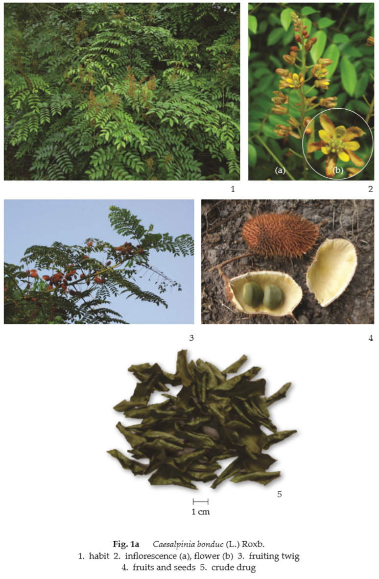
Nicker-Nut Leaf is the dried full-grown leaflets of Caesalpinia bonduc (L.) Roxb. [Guilandina bonduc Griseb., G. bonduccella L., Caesalpinia bonduccella (L.) Fleming] (Family Leguminosae), Herbarium Specimen Number: DMSC 1090, Crude Drug Number: DMSc 0277.
Constituents Nicker-Nut Leaf contains a bitter substance, waxy, and alcoholic substances.
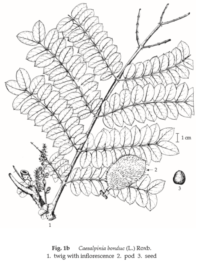
Description of the plant (Figs. 1a, 1b) Climber armed with straight or recurved prickles. Stipules, pinnate, consisting of 3 to 5 leaflets, up to 2 cm long. Leaves bipinnately compound, rachis 30 to 50 cm long; pinnate 3 to 9 pairs, with a pair of hooked stipulary spines at the base; leaflets, dark green, 8 to 12 pairs, opposite or subopposite, ovate- lanceolate, 2 to 4 cm long, 1 to 2 cm wide, acute or round and mucronulate apex, margin entire, base unequal; petiolule 0.8 mm long, stipels of short hooked spines. Inflorescence supra-axillary raceme, about 6 cm long, sometimes branched; flower yellow; bract linear, 6 to 12 mm long, recurved, lately caducous; pedicel 4 to 6 mm long, pubescent, faintly jointed near the top; calyx tube short, segments 5, pubescent, subequal; petals 5, yellow, the upper one with a long claw and hairy inside; stamens 10, free, filament hairy; ovary about 1 mm in diameter, stalked, hairy and bristly. Pod stalked above the receptacle (0.5 to 1 cm long), elliptic or oblong in outline, 5 to 8 cm long, 3 to 5 cm wide, set with hairy 7 to 9 mm long bristles. Seeds 2, subglobular, 15 to 20 mm in diameter, greyish.
Description
Macroscopical (Fig. 1a) Nicker-Nut Leaf occurs as a mixture of entire and broken brownish green leaflets and frequently rachillae of the compound leaf. Leaflet short, petiolule, lamina inequilaterally ovate-lanceolate, 2 to 4 cm long, 1 to 2 cm wide, acute or rounded and mucronulate apex, margin entire and base unequal, the upper surface brownish green, the lower surface pale greyish green and showing prominent midrib.
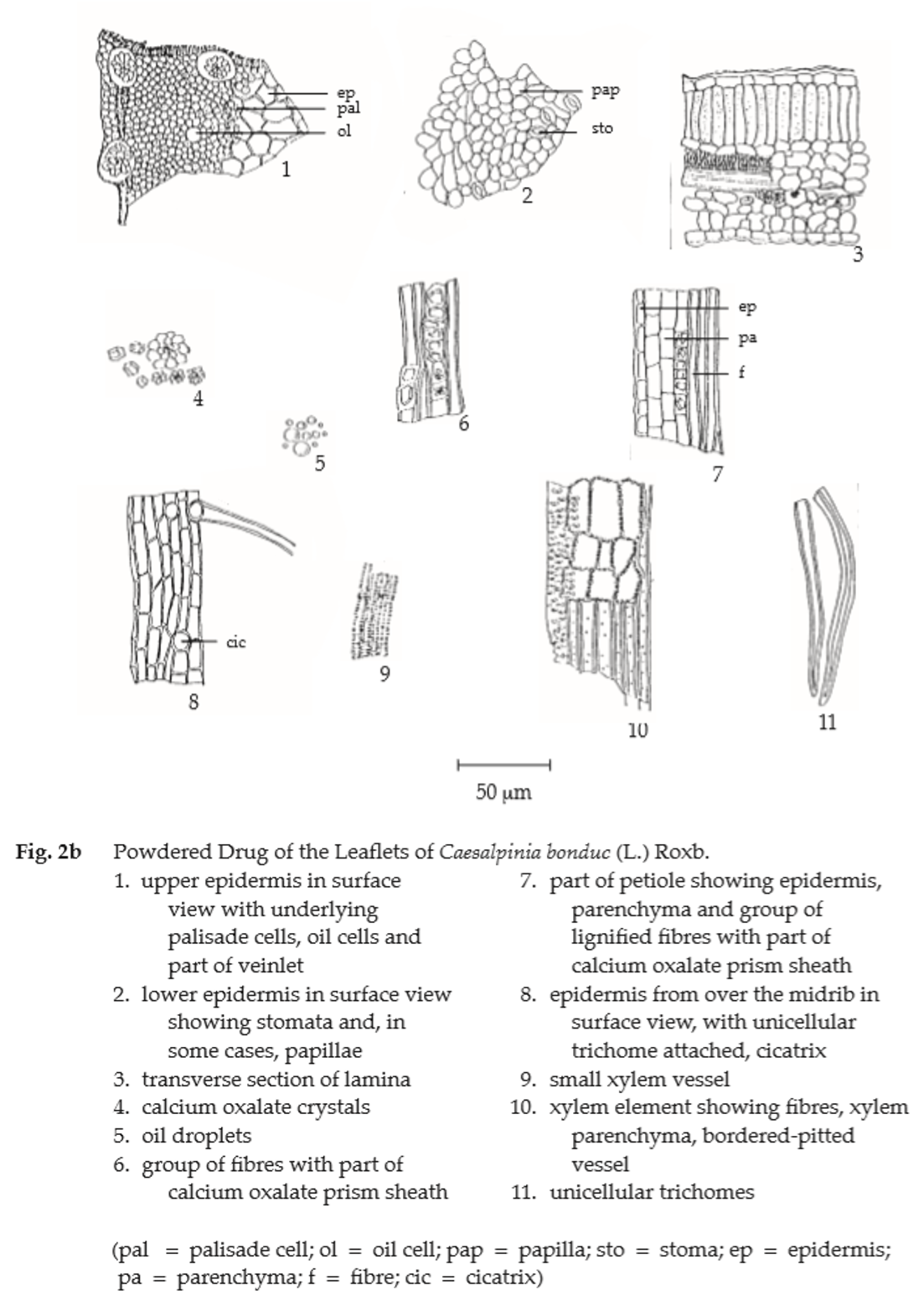
Microscopical (Figs. 2a, 2b) Transverse section of the leaflet shows upper epidermis, a single layer of cuticularized rectangular cells. Mesophyll, consisting of a single layer of palisade parenchyma associated with oil cells and several layers of irregular-shaped spongy parenchyma rich in chloroplastids and some oil cells near the lower epidermis. Small vascular bundles with calcium oxalate prism sheaths scatter in this parenchyma region. Lower epidermis, a single layer of cuticularized rectangular cells. Transverse section through the midrib of the lamina shows several layers of collenchyma underneath the epidermis, collateral vascular bundle in the centre and the long unicellular non-glandular trichomes on upper and lower epidermides.
In surface view, epidermides of the upper and lower surfaces are wavy-walled polygonal cells with anomocytic stomata in the lower epidermis.
Nicker-Nut Leaf in powder possesses the diagnostic microscopical characters of the unground drug.

Packaging and storage Nicker-Nut Leaf shall be kept in well-closed containers, protected from light, and stored in a dry place.
Identification
A. To 500 mg of the sample, in powder, add 2 mL of acetic anhydride, warm on a water-bath for about 2 minutes, shake, and filter. Slowly add 1 mL of sulfuric acid to the filtrate to form a layer: a red ring forms at the zone of contact.
B. Boil 500 mg of the sample, in powder, with 10 ml of water and filter. To 2 mL of the filtrate, add 1 drop of a 5 per cent w/v solution of iron(III) chloride: a brownish green precipitate is produced.
C. Reflux 500 mg of the sample, in powder, with 10 mL of methanol on a water-bath for 20 minutes and filter. To 2 mL of the filtrate, add 2 to 3 pieces of magnesium ribbon and 2 to 3 drops of hydrochloric acid, and warm on a water-bath: a brownish red colour develops.
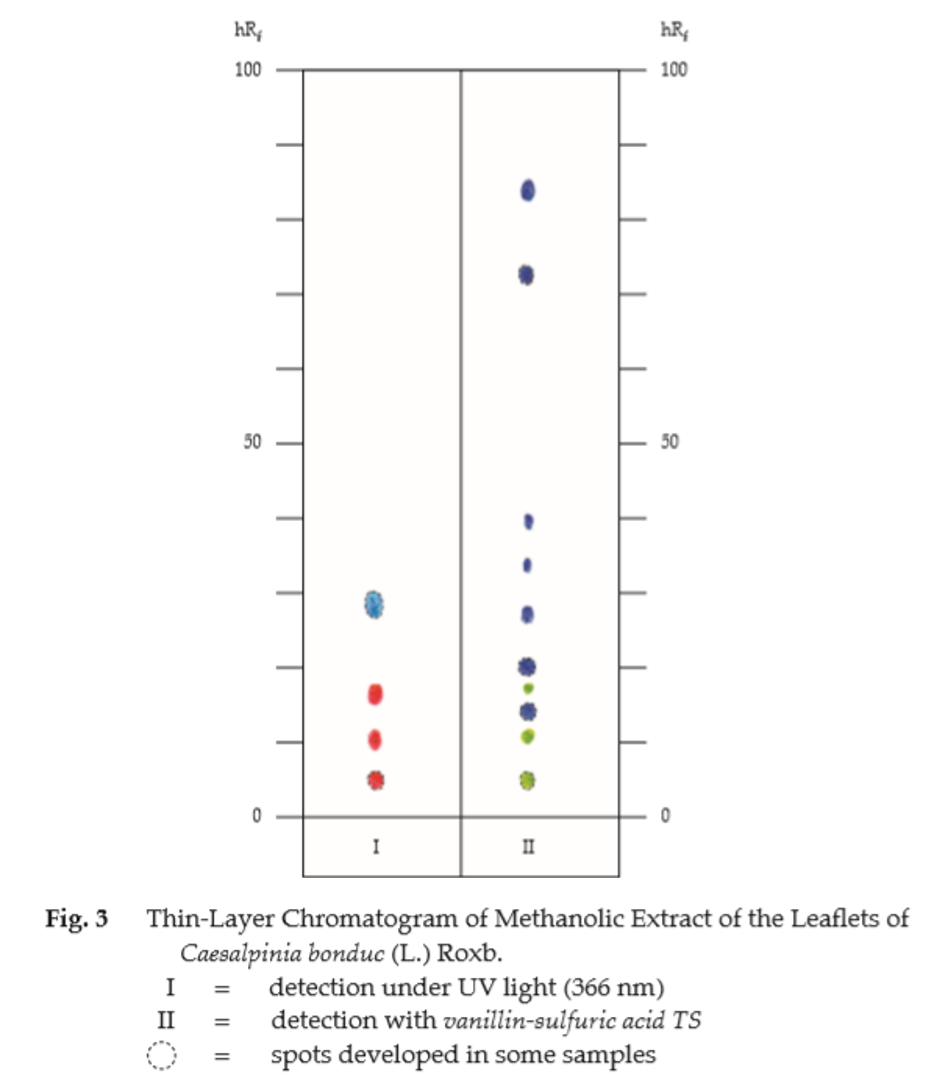
D. Carry out the test as described in the “Thin-Layer Chromatography” (Appendix 3.1), using silica gel G as the coating substance and a mixture of 85 volumes of hexane and 15 volumes of ethyl acetate as the mobile phase. Apply to the plate, 10 μL of the test solution prepared by refluxing 2 g of the sample, in powder, with 20 mL of methanol on a water-bath for about 30 minutes and filtering. After removal of the plate, allow it to dry in air and examine under ultraviolet light (366 nm); three red and one blue fluorescent spots appear. Spray the plate with vanillin-sulfuric acid TS and heat at 120° for 5 to 10 minutes; several spots of different colours are observed (Table 1); see also Fig. 3.
Table 1 hRf Values of Components in Methanolic Extract of the Leaflets of Caesalpinia bonduc (L.) Roxb.
| Spot | hRf Value | Detection | |
| UV 366 | Vanillin-Sulfuric Acid TS | ||
| 1 2 3 4 5 6 7 8 9 10 11 |
3-6 7-11 9-12 13-18 17-19 24-30 27-29 32-39 38-45 67-77 77-91 |
red red - red - - blue - - - - |
greenish grey greenish grey violet greenish grey violet violet - violet violet violet violet |
Loss on drying Not more than 11.0 per cent w/w after drying at 105° to constant weight (Appendix 4.15).
Acid-insoluble ash Not more than 1.5 per cent w/w (Appendix 7.6).
Sulfated ash Not more than 15.0 per cent w/w (Appendix 5.3).
Ethanol-soluble extractive Not less than 10.0 per cent w/w (Appendix 7.12).
Water-soluble extractive Not less than 25.0 per cent w/w (Appendix 7.12).
Chloroform-soluble extractive Not less than 6.0 per cent w/w (Appendix 7.12H).