ตำรามาตรฐานยาสมุนไพรไทย
Thai Herbal Pharmacopoeia
สำนักยาและวัตถุเสพติด กรมวิทยาศาสตร์การแพทย์ กระทรวงสาธารณสุข
Bureau of Drug and Narcotic, Department of Medical Sciences, Ministry of Public Healthสำนักยาและวัตถุเสพติด กรมวิทยาศาสตร์การแพทย์ กระทรวงสาธารณสุข
Bureau of Drug and Narcotic, Department of Medical Sciences, Ministry of Public Health(Tinospora crispa (L.) Hook.f. & Thomson)
(Nelumbo nucifera Gaertn.)
(Centella asiatica (L.) Urb.)
(Centella Dry Extract)
(Centella Cream)
(Mesua ferrea L.)
(Piper sarmentosum Roxb.)
(Piper sarmentosum Roxb.)
(Pterocarpus santalinus L. f.)
(Santalum album L.)
(Senna tora (L.) Roxb.)
(Senna alata (L.) Roxb.)
(Senna Alata Tea)
(Piper retrofractum Vahl)
(Myristica fragrans Houtt)
(Andrographis paniculata (Burm. f.) Nees)
(Andrographis Capsules)
(Allium ascalonicum L.)
(Ocimum tenuiflorum L.)
(Curcuma longa L.)
(Turmeric Capsules)
(Turmeric Dry Extract)
(Turmeric Dry Extract Capsules)
(Arcangelisia flava (L.) Merr.)
(Curcuma sp.)
Harrisonia perforata (Blanco) Merr.
(Aristolochia pierrei Lecomte)
(Zingiber officinale Roscoe)
(Ginger Capsules)
(Ginger Tea)
(Cassia fistula L.)
(Nardostachys jatamansi (D. Don) DC.)
(Angelica sinensis (Oliv.) Diels)
Artemisia annua L.
(Ligusticum sinense Oliv. cv. Chuanxiong)
(Neopicrorhiza scrophulariiflora Pennell)
(Atractylodes lancea (Thunb.) DC.)
(Aucklandia lappa Decne)
(Terminalia chebula Retz.)
(Angelica dahurica (Hoffm.) Benth. & Hook. f. ex Franch. & Sav. var. dahurica)
(Kaempferia parviflora Wall. ex Baker)
(Hibiscus sabdariffa L.)
(Roselle Tea)
(Allium sativum L.)
(Zingiber zerumbet (L.) Sm.)
(Wurfbainia testacea (Ridl.) Škorničk.& A. D. Poulsen)
(Cannabis sativa L.)
(Myristica fragrans Houtt)
(Dracaena cochinchinensis (Lour.) S. C. Chen)
(Ficus racemosa L.)
(Hyptis suaveolens (L.) Poit.)
Clerodendrum indicum (L.) Kuntze
(Phyllanthus emblica L.)
(Citrus hystrix DC.)
(Citrus hystrix DC.)
(Areca catechu L.)
(Momordica charantia L.)
Moringa oleifera Lam.
(Aegle marmelos (L.) Corrêa)
(Solanum trilobatum L.)
(Morus alba L.)
Gynostemma pentaphyllum(Thunb.)
Makino
(Clinacanthus nutans (Burm. f.) Lindau)
(Cissus quadrangularis L.)
(Mimusops elengi L.)
(Zingiber montanum (J. König) Link. ex A. Dietr.)
(Piper betle L.)
(Capsicum annuum L.)
(Capsicum Oleoresin)
(Capsicum Gel)
(Piper nigrum L.)
(Piper nigrum L.)
(Eurycoma longifolia Jack)
(Thunbergia laurifolia Lindl.)
(Piper wallichii (Miq.) Hand.-Mazz.)
Senna garrettiana (Craib) H. S. Irwin & Barneby
(Terminalia bellirica (Gaertn.) Roxb.)
(Terminalia chebula Retz.)
(Caesalpinia bonduc (L.) H. Roxb.)
(Tarlmounia elliptica (DC.) H. Rob., S. C. Keeley, Skvaria & R. Chan)
(Hog Creeper Vine Dry Extract Capsiles)
(Hog Creeper Vine Dry Extract)
(Brachypterum scandens (Roxb.) Miq.)
(Lepidium sativum L.)
(Nigella sativa L.)
(Cuminum cyminum L.)
(Foeniculum vulgare Mill.)
(Plantago ovata Forssk.)
(Pimpinella anisum L.)
(Carum carvi L.)
(Anethum graveolens L.)
(Trachyspermum ammi (L.) Sprague)
Albizia procera (Roxb.) Benth.
(Acorus calamus L.)
(Tiliacora triandra (Colebr.) Diels)
Cyanthillium cinereum (L.) H. Rob.
(Orthosiphon aristatus (Blume) Miq.)
Murdannia loriformis (Hassk.) R. S. Rao & Kammathy
(Capparis micracantha DC.)
(Chrysopogon zizanioides (L.) Roberty)
(Cyperus rotundus L.)
(Cannabis sativa L.)
(Syzygium aromaticum (L.) Merr. & L. M. Perry)
(Boesenbergia rotunda (L.) Mansf.)
(Acanthus ebracteatus Vahl)
(Acanthus ilicifolius L.)
(Kaempferia galanga L.)
(Curcuma comosa Roxb.)
Betula alnoides Buch.-Ham. ex D. Don
Cannabis sativa L.
Carthamus tinctorius L
Mitragyna speciosa (Korth.) Havil
Mallotus repandus (Rottler) Müll. Arg
Azadirachta indica A. Juss. var. siamensis Valeton
Azadirachta indica A. Juss. var. siamensis Valeton
Punica granatum L.
Rhinacanthus nasutus (L.) Kurz
Baliospermum solanifolium (Burm.) Suresh
Curcuma aeruginosa Roxb
Boesenbergia kingii Mood & L. M. Prince
Senegalia rugata (Lam.) Britton & Rose
Acacia concinna (Willd.) DC.
Senegalia rugata (Lam.) Britton & Rose
Acacia concinna (Willd.) DC.
Senna alexandriana Mill. var. alexandriana
Cassia acutifolia Delile, Cassia angustifolia Vahl
Butea superba Roxb. ex Willd.
[Plaso superba (Roxb. ex Willd.) Kuntze, Rudolphia superba (Roxb. ex Willd.) Poir.
Pueraria candollei Graham
ex Benth. var. mirifica (Airy Shaw & Suvat.) Niyomdham
Streblus asper Lour.
Suregada multiflora (A. Juss.) Baill. (Gelonium
multiflorum A. Juss.
Plumbago zeylanica L.
Plumbago indica L.
Biancaea sappan (L.) Tod.
Ziziphus attopensis Pierre
Streblus asper Lour.
Justicia gendarussa Burm. f.
Enhalus acoroides (L. f.) Royle
Bridelia ovata Decne.
Tamarindus indica L.
Citrus × aurantiifolia (Christm.) Swingle
Garcinia mangostana L.
Blumea balsamifera (L.) DC
Persicaria odorata (Lour.) Soják
Zingiber montanum (J. König) Link ex A. Dietr.
Mammea siamensis (Miq.) T. Anderson
Citrus maxima (Burm.) Merr.
Citrus × aurantium L. ‘Som Sa'
Punica granatum L.
Rhinacanthus nasutus (L.) Kurz
Betel Leaf is the dried leaf of Piper betle L. (Family Piperaceae), Herbarium Specimen Number: DMSC 902.
Constituents Betel Leaf contains volatile oil comprising cadinene, carvacrol, caryophyllene, chavibetol, chavicol, 1,8-cineole, estragole, eugenol, terpinyl acetate, etc. It also contains sitosterols, stigmasterol, vitamin C, oxalic acid, d(+)malic acid, n-hentriacontane, n-pentatriacontane, amino acids, pyridine alkaloids, tannins, minerals, etc.
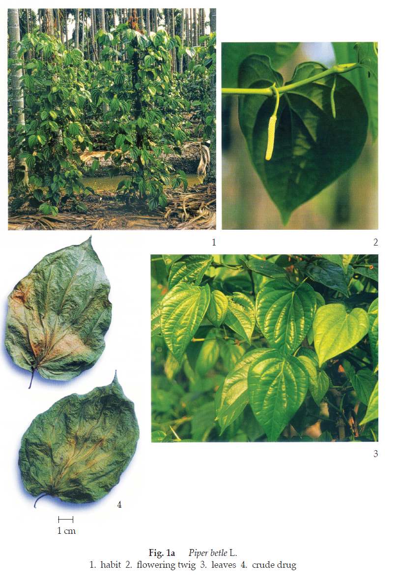
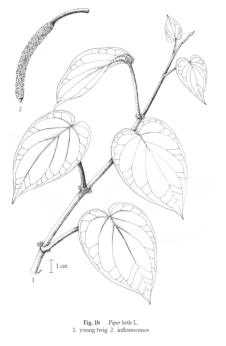
Description of the plant (Figs. 1a, 1b) Woody climber with swollen nodes, up to 15 m tall, dimorphic branching; orthotropic vegetative branches with adventitious roots; plagiotropic axillary fruiting branches without roots. Leaves simple, circular, ovate or ovate-oblong, 5 to 18 cm long, 3 to 12 cm wide; surface glabrous or with very short, thick white hairs, apex acuminate, base cordate or obliquely obtuse, margin entire, veins prominent on lower surface; petiole cylindrical, 1.2 to 2.5 cm long. Inflorescence in drooping, dense axillary spike, consisting of male and female flowers. Male spike 2.5 to 12 cm long; peduncle 1.5 to 3 cm long; stamens 2, very short. Female spike 2.5 to 12 cm long; peduncle 2.5 to 6 cm long; stigmas 3 to 5. Fruit berry, round with glabrous apex. Seed suborbicular, 3.5 to 5 mm long.
Description Odour, aromatic; taste, pungent.
Macroscopical (Fig. 1a) Leaves, simple, circular, ovate or ovate-oblong, 5 to 18 cm long, 3 to 12 cm wide; acute acuminate; base cordate or obliquely obtuse; upper surface smooth, dull brown; lower surface dull, lighter colour.
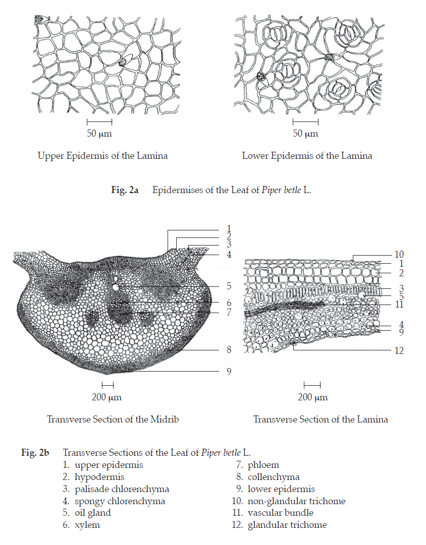
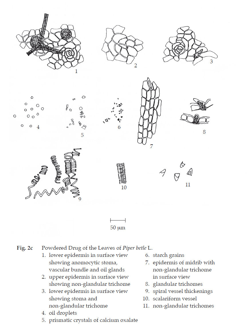
Microscopical (Figs. 2a, 2b, 2c) Transverse section of the leaf shows upper epidermis, a layer of quadrangular cells, polygonal and straight-walled in surface view; cuticle thick; stoma absent with few glandular and non-glandular trichomes. Non-glandular trichome consists of a single short conical thick-walled cell tapering to the apex, and glandular trichome with a unicellular round head. Lower epidermis, almost similar to those of upper epidermis, polygonal and rather wavy walls in surface view; anomocytic stomata, numerous. Hypodermis, two to three layers of cells, some of which being oil glands. Mesophyll consists of a layer of palisade cells and a few layers of irregular spongy parenchyma containing abundant chloroplastids; oil glands, numerous. Vascular bundle, collateral, a few layers of collenchyma beneath upper and lower epidermises, schizogenous oil cavities, crystals and some starch granules in the midrib.
Betel Leaf in powder possesses the diagnostic microscopical characters of the unground drug.
Packaging and storage Betel Leaf shall be kept in well-closed containers, protected from light, and stored in a dry place.
Identification
A. Macerate 5 g of the sample, in powder, with 50 mL of water for 15 minutes. Distil for about 3 to 4 hours. Extract the distillate with three successive 25-mL, 20-mL and 15-mL portions of dichloromethane. Filter over anhydrous sodium sulfate, evaporate to dryness. Add 1 mL of chloroform and 1 mL of acetic anhydride to the residue. Slowly add 1 mL of sulfuric acid to form a layer: a violet brown colour forms at the zone of contact.
B. Macerate 500 mg of the sample, in powder, with 10 mL of ethanol for 5 minutes and filter. Shake 5 mL of the filtrate with 200 mg of decolorizing charcoal, filter and add 1 drop of iron(III) chloride TS to 1 mL of the filtrate: a green colour is produced.
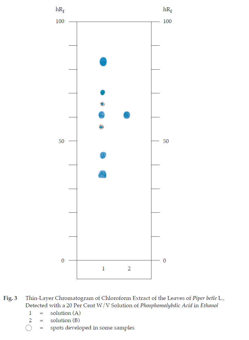
C. Carry out the test as described in the “Thin-Layer Chromatography” (Appendix 3.1), using silica gel GF254 as the coating substance and a mixture of 75 volumes of toluene and 25 volumes of ethyl acetate as the mobile phase. Apply separately to the plate, 1 μL each of the following solutions. Prepare solution (A) by macerating 2 g of the sample, in powder, with 20 mL of chloroform for 24 hours and filtering. Evaporate the filtrate under reduced pressure at 40° until dryness, and dissolve the residue in 2 mL of chloroform. For solution (B), dilute 5 μL of eugenol with chloroform to make 1 mL. After removal of the plate, allow it to dry in air, spray the plate with a 20 per cent w/v solution of phosphomolybdic acid in ethanol and heat at 105° for 5 minutes; a blue spot due to eugenol (hRf value 60 to 62) and other four to six spots of blue and greenish blue colours are observed (Table 1); see also Fig. 3. When treated with ammonia vapour; the yellow background of the layer turns white.
Table 1 hRf Values of Components in Chloroform Extract of the Leaves of Piper betle L.
| Spot | hRf Value | Detection |
| 20 Per Cent W/V Solution of Phosphomolybdic Acid in Ethanol |
||
| 1 2 3 4* 5 6 7 |
33-38 43-45 55-57 60-62 63-69 66-67 78-88 |
blue blue greenish blue blue greenish blue greenish blue blue |
*eugenol
Water Not more than 8.0 per cent v/w (Azeotropic Distillation Method, Appendix 4.12).
Foreign matter Not more than 2.0 per cent w/w (Appendix 7.2).
Acid-insoluble ash Not more than 7.0 per cent w/w (Appendix 7.6).
Total ash Not more than 14.0 per cent w/w (Appendix 7.7).
Ethanol-soluble extractive Not less than 4.0 per cent w/w (Appendix 7.12).
Water-soluble extractive Not less than 14.0 per cent w/w (Appendix 7.12).