ตำรามาตรฐานยาสมุนไพรไทย
Thai Herbal Pharmacopoeia
สำนักยาและวัตถุเสพติด กรมวิทยาศาสตร์การแพทย์ กระทรวงสาธารณสุข
Bureau of Drug and Narcotic, Department of Medical Sciences, Ministry of Public Healthสำนักยาและวัตถุเสพติด กรมวิทยาศาสตร์การแพทย์ กระทรวงสาธารณสุข
Bureau of Drug and Narcotic, Department of Medical Sciences, Ministry of Public Health(Tinospora crispa (L.) Hook.f. & Thomson)
(Nelumbo nucifera Gaertn.)
(Centella asiatica (L.) Urb.)
(Centella Dry Extract)
(Centella Cream)
(Mesua ferrea L.)
(Piper sarmentosum Roxb.)
(Piper sarmentosum Roxb.)
(Pterocarpus santalinus L. f.)
(Santalum album L.)
(Senna tora (L.) Roxb.)
(Senna alata (L.) Roxb.)
(Senna Alata Tea)
(Piper retrofractum Vahl)
(Myristica fragrans Houtt)
(Andrographis paniculata (Burm. f.) Nees)
(Andrographis Capsules)
(Allium ascalonicum L.)
(Ocimum tenuiflorum L.)
(Curcuma longa L.)
(Turmeric Capsules)
(Turmeric Dry Extract)
(Turmeric Dry Extract Capsules)
(Arcangelisia flava (L.) Merr.)
(Curcuma sp.)
Harrisonia perforata (Blanco) Merr.
(Aristolochia pierrei Lecomte)
(Zingiber officinale Roscoe)
(Ginger Capsules)
(Ginger Tea)
(Cassia fistula L.)
(Nardostachys jatamansi (D. Don) DC.)
(Angelica sinensis (Oliv.) Diels)
Artemisia annua L.
(Ligusticum sinense Oliv. cv. Chuanxiong)
(Neopicrorhiza scrophulariiflora Pennell)
(Atractylodes lancea (Thunb.) DC.)
(Aucklandia lappa Decne)
(Terminalia chebula Retz.)
(Angelica dahurica (Hoffm.) Benth. & Hook. f. ex Franch. & Sav. var. dahurica)
(Kaempferia parviflora Wall. ex Baker)
(Hibiscus sabdariffa L.)
(Roselle Tea)
(Allium sativum L.)
(Zingiber zerumbet (L.) Sm.)
(Wurfbainia testacea (Ridl.) Škorničk.& A. D. Poulsen)
(Cannabis sativa L.)
(Myristica fragrans Houtt)
(Dracaena cochinchinensis (Lour.) S. C. Chen)
(Ficus racemosa L.)
(Hyptis suaveolens (L.) Poit.)
Clerodendrum indicum (L.) Kuntze
(Phyllanthus emblica L.)
(Citrus hystrix DC.)
(Citrus hystrix DC.)
(Areca catechu L.)
(Momordica charantia L.)
Moringa oleifera Lam.
(Aegle marmelos (L.) Corrêa)
(Solanum trilobatum L.)
(Morus alba L.)
Gynostemma pentaphyllum(Thunb.)
Makino
(Clinacanthus nutans (Burm. f.) Lindau)
(Cissus quadrangularis L.)
(Mimusops elengi L.)
(Zingiber montanum (J. König) Link. ex A. Dietr.)
(Piper betle L.)
(Capsicum annuum L.)
(Capsicum Oleoresin)
(Capsicum Gel)
(Piper nigrum L.)
(Piper nigrum L.)
(Eurycoma longifolia Jack)
(Thunbergia laurifolia Lindl.)
(Piper wallichii (Miq.) Hand.-Mazz.)
Senna garrettiana (Craib) H. S. Irwin & Barneby
(Terminalia bellirica (Gaertn.) Roxb.)
(Terminalia chebula Retz.)
(Caesalpinia bonduc (L.) H. Roxb.)
(Tarlmounia elliptica (DC.) H. Rob., S. C. Keeley, Skvaria & R. Chan)
(Hog Creeper Vine Dry Extract Capsiles)
(Hog Creeper Vine Dry Extract)
(Brachypterum scandens (Roxb.) Miq.)
(Lepidium sativum L.)
(Nigella sativa L.)
(Cuminum cyminum L.)
(Foeniculum vulgare Mill.)
(Plantago ovata Forssk.)
(Pimpinella anisum L.)
(Carum carvi L.)
(Anethum graveolens L.)
(Trachyspermum ammi (L.) Sprague)
Albizia procera (Roxb.) Benth.
(Acorus calamus L.)
(Tiliacora triandra (Colebr.) Diels)
Cyanthillium cinereum (L.) H. Rob.
(Orthosiphon aristatus (Blume) Miq.)
Murdannia loriformis (Hassk.) R. S. Rao & Kammathy
(Capparis micracantha DC.)
(Chrysopogon zizanioides (L.) Roberty)
(Cyperus rotundus L.)
(Cannabis sativa L.)
(Syzygium aromaticum (L.) Merr. & L. M. Perry)
(Boesenbergia rotunda (L.) Mansf.)
(Acanthus ebracteatus Vahl)
(Acanthus ilicifolius L.)
(Kaempferia galanga L.)
(Curcuma comosa Roxb.)
Betula alnoides Buch.-Ham. ex D. Don
Cannabis sativa L.
Carthamus tinctorius L
Mitragyna speciosa (Korth.) Havil
Mallotus repandus (Rottler) Müll. Arg
Azadirachta indica A. Juss. var. siamensis Valeton
Azadirachta indica A. Juss. var. siamensis Valeton
Punica granatum L.
Rhinacanthus nasutus (L.) Kurz
Baliospermum solanifolium (Burm.) Suresh
Curcuma aeruginosa Roxb
Boesenbergia kingii Mood & L. M. Prince
Senegalia rugata (Lam.) Britton & Rose
Acacia concinna (Willd.) DC.
Senegalia rugata (Lam.) Britton & Rose
Acacia concinna (Willd.) DC.
Senna alexandriana Mill. var. alexandriana
Cassia acutifolia Delile, Cassia angustifolia Vahl
Butea superba Roxb. ex Willd.
[Plaso superba (Roxb. ex Willd.) Kuntze, Rudolphia superba (Roxb. ex Willd.) Poir.
Pueraria candollei Graham
ex Benth. var. mirifica (Airy Shaw & Suvat.) Niyomdham
Streblus asper Lour.
Suregada multiflora (A. Juss.) Baill. (Gelonium
multiflorum A. Juss.
Plumbago zeylanica L.
Plumbago indica L.
Biancaea sappan (L.) Tod.
Ziziphus attopensis Pierre
Streblus asper Lour.
Justicia gendarussa Burm. f.
Enhalus acoroides (L. f.) Royle
Bridelia ovata Decne.
Tamarindus indica L.
Citrus × aurantiifolia (Christm.) Swingle
Garcinia mangostana L.
Blumea balsamifera (L.) DC
Persicaria odorata (Lour.) Soják
Zingiber montanum (J. König) Link ex A. Dietr.
Mammea siamensis (Miq.) T. Anderson
Citrus maxima (Burm.) Merr.
Citrus × aurantium L. ‘Som Sa’
Punica granatum L.
Rhinacanthus nasutus (L.) Kurz
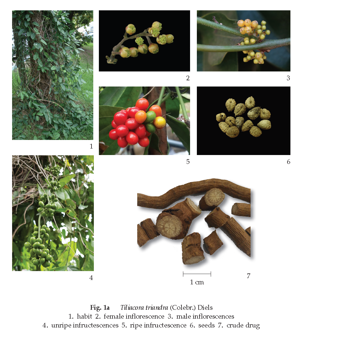
Tiliacora Triandra Root is the dried root of Tiliacora triandra (Colebr.) Diels (Coccolus triandrus Colebr., Limacia triandra (Colebr.) Hook. f.) (Family Menispermaceae), Herbarium Specimen Number: DMSC 5210, Crude Drug Number: DMSc 1071.
Constituents Tiliacora Triandra Root contains bisbenzylisoquinoline alkaloids (e.g., 2’-nortiliacorinine, tiliacorine, tiliacorinine, yanangcorinine).
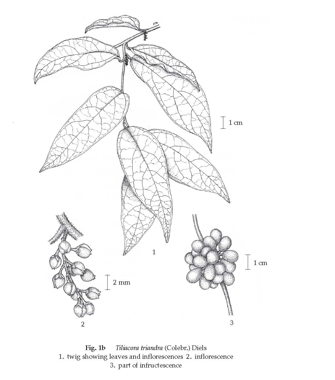
Description of the plant (Figs. 1a, 1b) Climber or straggling shrub; stem usually slender, glabrous or pubescent, striate. Leaves simple, alternate, ovate, elliptic or lanceolate, 5 to 11(–17) cm long, 2 to 6(–10) cm wide, apex obtuse, acute or acuminate, base attenuate, obtuse or subcordate, margin entire, glabrous, papyraceous, pinnately nerved, often with steeply ascending basal nerves, seemingly subpalmately nerved, midrib of lower surface rugulose near base; petiole 0.5 to 2 cm long, rugulose. Inflorescence paniculate, 1- to few-flowered, axillary or cauliflorous, 2 to 8(–17) cm long; peduncle about 5 mm long. Male flower yellowish, about 2 mm in diameter; sepals 6, 2-seriate, 3 outer ones smaller, 3 inner ones broadly elliptic, about 2 mm long, subglabrous; petals 3 or 6, minute glabrous; stamens 3, clavate, about 2 mm long. Female flower reddish; sepal as in male flower; petals 6, oblongelliptic, about 1 mm long; carpels 8 or 9, less than 1 mm long, borne on short gynophore, glabrous, stigma sessile; staminode absent. Fruit drupe, obovoid, 0.7 to 1 cm long, 6 to 7 mm wide, subcompressed, glabrous, green when young becoming yel lowish or reddish when ripe; stalk 3 to 4 mm long; endocarp transversely and irregularly ridged. Seed 1, horseshoe-shaped, curved, rugulose.
Description Odourless; taste, bitter.
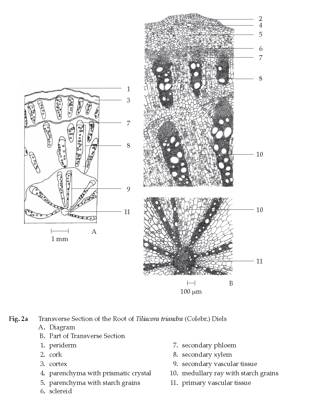
Macroscopical (Fig. 1a) Transverse slices of roots; bark thin, brown; sectional surface of wood yellowish to light brown, with several successive and distinct concentric and radiate zones.
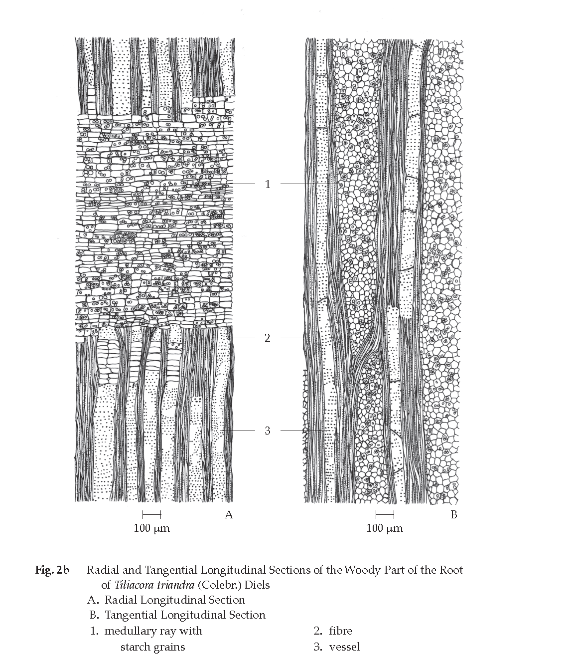
Microscopical (Figs. 2a, 2b, 2c) Transverse section of the root shows periderm, cortex and vascular tissue. Periderm, several layers of thin-walled rectangular cork cells. Cortex, polygonal shape of thin-walled parenchyma cells, some containing starch grains or prismatic crystals, and several layers of thick-walled sclereids in the inner zone. Vascular tissue, primary and secondary vascular tissues; primary vascular tissue, metaphloem and metaxylem; secondary vascular tissue, anomalous type, radially arranged, some containing secondary phloem, secondary xylem and broad zone of ray cells, some of which contain starch grains.
Radial and tangential longitudinal sections of the woody part of the root illustrate medullary ray cells with starch grains, bordered-pitted vessels and bundles of fibres.
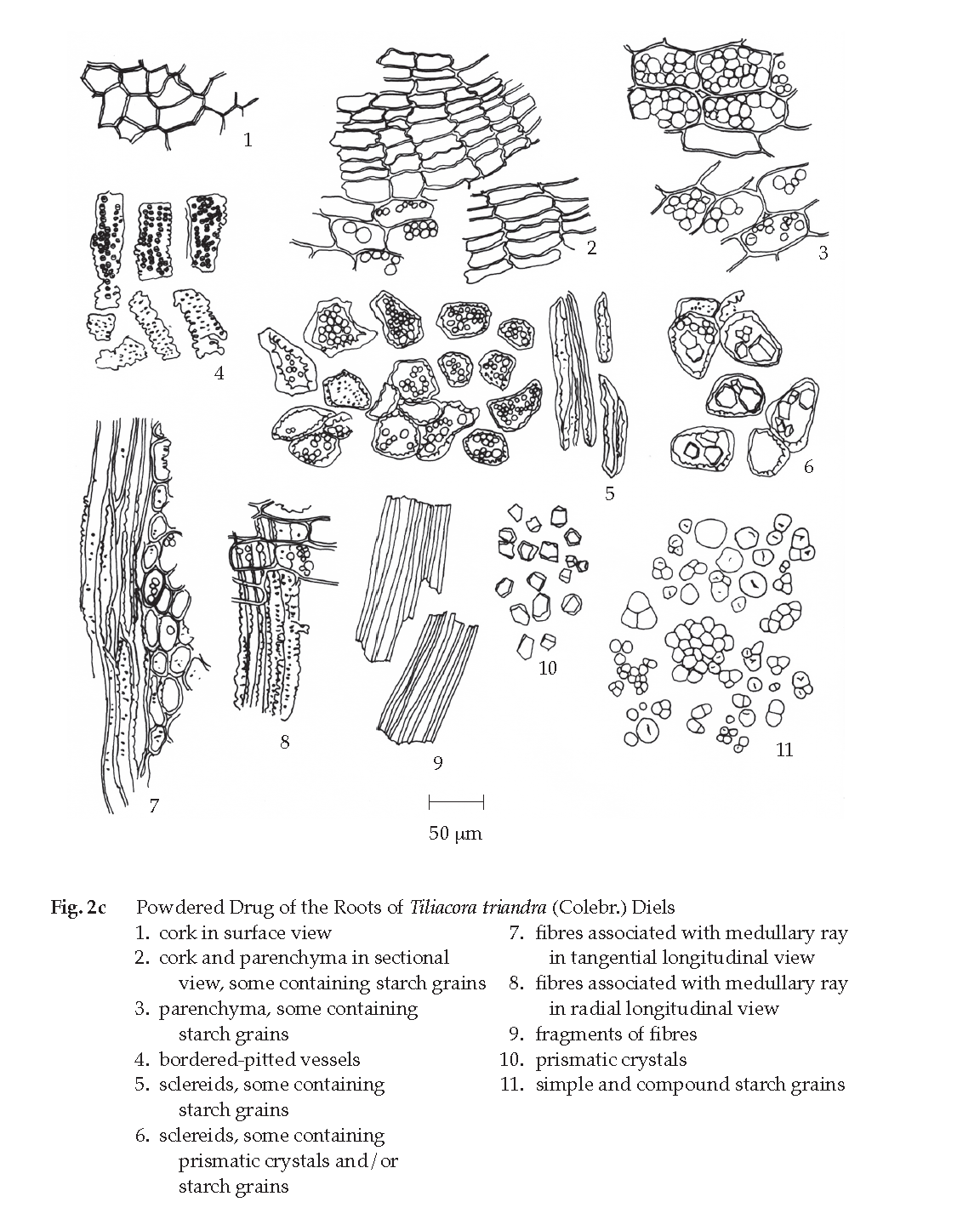
Tiliacora Triandra Root in powder possesses the diagnostic microscopical characters of the unground drug. Various shapes of sclereids, some containing prismatic crystals, simple and compound starch grains, scattered prismatic crystals and thin-walled corks are frequently observed.
Packaging and storage Tiliacora Triandra Root shall be kept in well-closed containers, protected from light, and stored in a dry place.
Additional information
1. In Thai herbal markets, the adulteration of tiliacora triandra root with stems, branches, etc., may be found.
2. Risk-benefit should be considered when consuming since safety data in humans are not available at the present time.
Identification
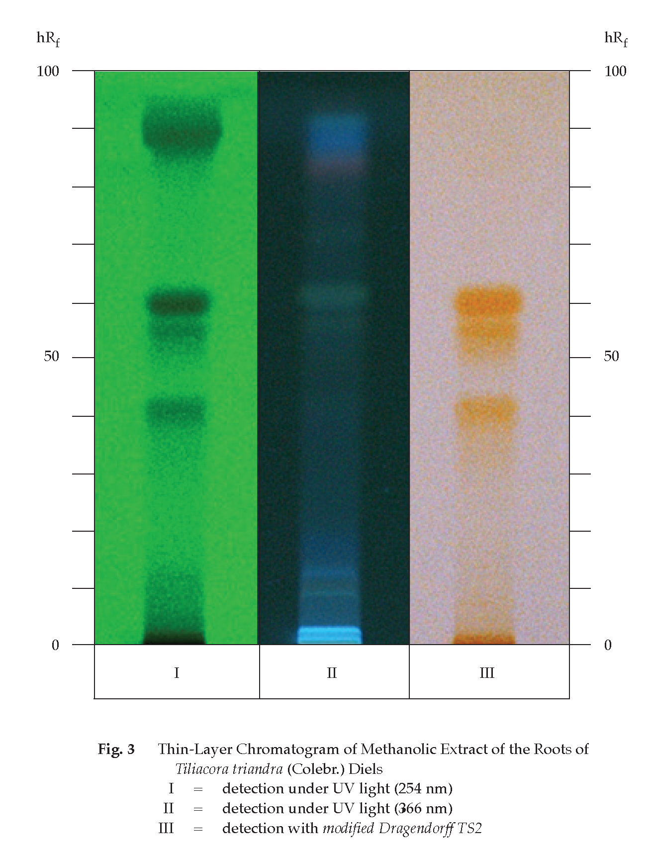
A. Macerate 200 mg of the sample, in fine powder, with 4 mL of chloroform for 5 minutes and filter. To 2 mL of the filtrate, add a few drops of modified Dragendorff TS2 and shake well: an orange precipitate is produced.
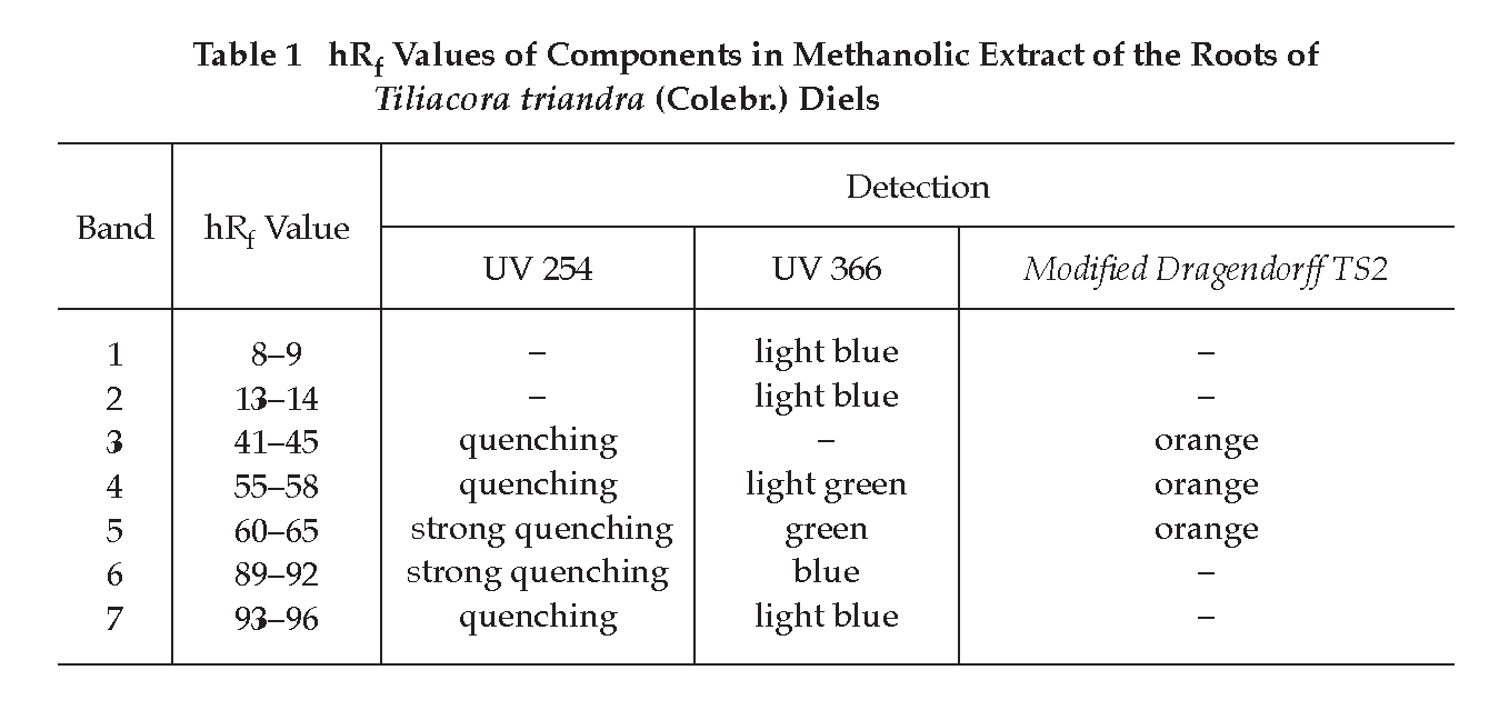
B. Carry out the test as described in the “Thin-Layer Chromatography” (Appendix 3.1), using silica gel GF254 as the coating substance and a mixture of 70 volumes of chloroform and 30 volumes of ethanol as the mobile phase and allowing the solvent front to ascend 8 cm above the line of application. Apply to the plate as a band of 8 mm, 5 μL of the test solution prepared by macerating 1 g of the sample, in fine powder, with 10 mL of methanol for 15 minutes, filtering and evaporating the filtrate to dryness. Dissolve the residue in 2 mL of methanol. After removal of the plate, allow it to dry in air and examine under ultraviolet light (254 nm), marking the quenching bands. Examine the plate under ultraviolet light (366 nm) through the cut-off filter; several fluorescent bands of different colours are observed. Spray the plate with modified Dragendorff TS2 and immediately examine the plate; three orange bands are observed (Table 1); see also Fig. 3.
Loss on drying Not more than 9.0 per cent w/w after drying at 105° to constant weight (Appendix 4.15).
Foreign matter Not more than 2.0 per cent w/w (Appendix 7.2).
Total ash Not more than 8.0 per cent w/w (Appendix 7.7).
Ethanol-soluble extractive Not less than 4.0 per cent w/w (Appendix 7.12).
Water-soluble extractive Not less than 6.0 per cent w/w (Appendix 7.12).