ตำรามาตรฐานยาสมุนไพรไทย
Thai Herbal Pharmacopoeia
สำนักยาและวัตถุเสพติด กรมวิทยาศาสตร์การแพทย์ กระทรวงสาธารณสุข
Bureau of Drug and Narcotic, Department of Medical Sciences, Ministry of Public Healthสำนักยาและวัตถุเสพติด กรมวิทยาศาสตร์การแพทย์ กระทรวงสาธารณสุข
Bureau of Drug and Narcotic, Department of Medical Sciences, Ministry of Public Health(Tinospora crispa (L.) Hook.f. & Thomson)
(Nelumbo nucifera Gaertn.)
(Centella asiatica (L.) Urb.)
(Centella Dry Extract)
(Centella Cream)
(Mesua ferrea L.)
(Piper sarmentosum Roxb.)
(Piper sarmentosum Roxb.)
(Pterocarpus santalinus L. f.)
(Santalum album L.)
(Senna tora (L.) Roxb.)
(Senna alata (L.) Roxb.)
(Senna Alata Tea)
(Piper retrofractum Vahl)
(Myristica fragrans Houtt)
(Andrographis paniculata (Burm. f.) Nees)
(Andrographis Capsules)
(Allium ascalonicum L.)
(Ocimum tenuiflorum L.)
(Curcuma longa L.)
(Turmeric Capsules)
(Turmeric Dry Extract)
(Turmeric Dry Extract Capsules)
(Arcangelisia flava (L.) Merr.)
(Curcuma sp.)
Harrisonia perforata (Blanco) Merr.
(Aristolochia pierrei Lecomte)
(Zingiber officinale Roscoe)
(Ginger Capsules)
(Ginger Tea)
(Cassia fistula L.)
(Nardostachys jatamansi (D. Don) DC.)
(Angelica sinensis (Oliv.) Diels)
Artemisia annua L.
(Ligusticum sinense Oliv. cv. Chuanxiong)
(Neopicrorhiza scrophulariiflora Pennell)
(Atractylodes lancea (Thunb.) DC.)
(Aucklandia lappa Decne)
(Terminalia chebula Retz.)
(Angelica dahurica (Hoffm.) Benth. & Hook. f. ex Franch. & Sav. var. dahurica)
(Kaempferia parviflora Wall. ex Baker)
(Hibiscus sabdariffa L.)
(Roselle Tea)
(Allium sativum L.)
(Zingiber zerumbet (L.) Sm.)
(Wurfbainia testacea (Ridl.) Škorničk.& A. D. Poulsen)
(Cannabis sativa L.)
(Myristica fragrans Houtt)
(Dracaena cochinchinensis (Lour.) S. C. Chen)
(Ficus racemosa L.)
(Hyptis suaveolens (L.) Poit.)
Clerodendrum indicum (L.) Kuntze
(Phyllanthus emblica L.)
(Citrus hystrix DC.)
(Citrus hystrix DC.)
(Areca catechu L.)
(Momordica charantia L.)
Moringa oleifera Lam.
(Aegle marmelos (L.) Corrêa)
(Solanum trilobatum L.)
(Morus alba L.)
Gynostemma pentaphyllum(Thunb.)
Makino
(Clinacanthus nutans (Burm. f.) Lindau)
(Cissus quadrangularis L.)
(Mimusops elengi L.)
(Zingiber montanum (J. König) Link. ex A. Dietr.)
(Piper betle L.)
(Capsicum annuum L.)
(Capsicum Oleoresin)
(Capsicum Gel)
(Piper nigrum L.)
(Piper nigrum L.)
(Eurycoma longifolia Jack)
(Thunbergia laurifolia Lindl.)
(Piper wallichii (Miq.) Hand.-Mazz.)
Senna garrettiana (Craib) H. S. Irwin & Barneby
(Terminalia bellirica (Gaertn.) Roxb.)
(Terminalia chebula Retz.)
(Caesalpinia bonduc (L.) H. Roxb.)
(Tarlmounia elliptica (DC.) H. Rob., S. C. Keeley, Skvaria & R. Chan)
(Hog Creeper Vine Dry Extract Capsiles)
(Hog Creeper Vine Dry Extract)
(Brachypterum scandens (Roxb.) Miq.)
(Lepidium sativum L.)
(Nigella sativa L.)
(Cuminum cyminum L.)
(Foeniculum vulgare Mill.)
(Plantago ovata Forssk.)
(Pimpinella anisum L.)
(Carum carvi L.)
(Anethum graveolens L.)
(Trachyspermum ammi (L.) Sprague)
Albizia procera (Roxb.) Benth.
(Acorus calamus L.)
(Tiliacora triandra (Colebr.) Diels)
Cyanthillium cinereum (L.) H. Rob.
(Orthosiphon aristatus (Blume) Miq.)
Murdannia loriformis (Hassk.) R. S. Rao & Kammathy
(Capparis micracantha DC.)
(Chrysopogon zizanioides (L.) Roberty)
(Cyperus rotundus L.)
(Cannabis sativa L.)
(Syzygium aromaticum (L.) Merr. & L. M. Perry)
(Boesenbergia rotunda (L.) Mansf.)
(Acanthus ebracteatus Vahl)
(Acanthus ilicifolius L.)
(Kaempferia galanga L.)
(Curcuma comosa Roxb.)
Betula alnoides Buch.-Ham. ex D. Don
Cannabis sativa L.
Carthamus tinctorius L
Mitragyna speciosa (Korth.) Havil
Mallotus repandus (Rottler) Müll. Arg
Azadirachta indica A. Juss. var. siamensis Valeton
Azadirachta indica A. Juss. var. siamensis Valeton
Punica granatum L.
Rhinacanthus nasutus (L.) Kurz
Baliospermum solanifolium (Burm.) Suresh
Curcuma aeruginosa Roxb
Boesenbergia kingii Mood & L. M. Prince
Senegalia rugata (Lam.) Britton & Rose
Acacia concinna (Willd.) DC.
Senegalia rugata (Lam.) Britton & Rose
Acacia concinna (Willd.) DC.
Senna alexandriana Mill. var. alexandriana
Cassia acutifolia Delile, Cassia angustifolia Vahl
Butea superba Roxb. ex Willd.
[Plaso superba (Roxb. ex Willd.) Kuntze, Rudolphia superba (Roxb. ex Willd.) Poir.
Pueraria candollei Graham
ex Benth. var. mirifica (Airy Shaw & Suvat.) Niyomdham
Streblus asper Lour.
Suregada multiflora (A. Juss.) Baill. (Gelonium
multiflorum A. Juss.
Plumbago zeylanica L.
Plumbago indica L.
Biancaea sappan (L.) Tod.
Ziziphus attopensis Pierre
Streblus asper Lour.
Justicia gendarussa Burm. f.
Enhalus acoroides (L. f.) Royle
Bridelia ovata Decne.
Tamarindus indica L.
Citrus × aurantiifolia (Christm.) Swingle
Garcinia mangostana L.
Blumea balsamifera (L.) DC
Persicaria odorata (Lour.) Soják
Zingiber montanum (J. König) Link ex A. Dietr.
Mammea siamensis (Miq.) T. Anderson
Citrus maxima (Burm.) Merr.
Citrus × aurantium L. ‘Som Sa'
Punica granatum L.
Rhinacanthus nasutus (L.) Kurz
Siamese Neem Leaf is the dried leaflets of Azadirachta indica A. Juss. var. siamensis Valeton (Family Meliaceae), Herbarium Specimen Number: DMSC 5336, BK 070190, Crude Drug Number: DMSc 1230.
Constituents Siamese Neem Leaf contains tetranortriterpenoids (e.g., azadirachtin) and flavonoids (e.g., quercetin and rutin).
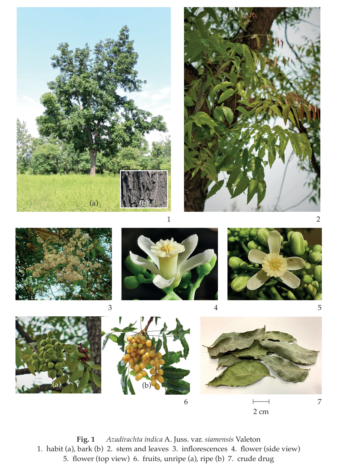
Description of plant (Fig. 1) Evergreen tree, up to 20 m tall; bark greyish brown, vertically striated. Leaves pinnately compound, spirally arranged, young leaves usually reddish; exstipulate; rachis 20 to 40 cm long; petiole 6 to 14 cm long, slender, swollen at base; leaflets 6 to 12 pairs, opposite or subopposite, leaflet at tip sometimes reduced; exstipellate; petiolule 4 to 7 mm long, slender, glabrous; lamina oblanceolate or broadly falcate, 6.5 to 12.5 cm long, 2.1 to 5.2 cm wide, apex acuminate, base oblique, margin serrate or serrate-undulate, coriaceous, glabrous, lateral nerves 8 to 13 pairs, slender, prominent, veinlets reticulate, fainted. Inflorescence panicle, axillary or terminal, up to 30 cm long, appearing before leaves. Flower white, bisexual, 1 to 1.5 cm in diameter; bracteole scaly; pedicel about 2 mm long; sepals 5, free, ovate; petals 5, free, obovate-oblong, hairy, imbricate in bud; stamens 10, connated into cylindrical staminal tube, about 6 mm long, terminally 10-lobed, glabrous outside, inside pubescent, anthers 10, attached at base of lobes inside, slightly exserted; ovary superior, globose, 3-loculed, ovules 2 per locule, placentation axile, style slender, elongate, stigma slightly 3-lobed. Fruit a drupe, oblong-ovoid, 1.5 to 2 cm long, 0.5 to 1 cm wide, green when young, yellow when ripe, shiny. Seeds 1 or 2.
Description Odour, green, characteristic; taste, bitter
Macroscopical (Fig. 1) A mixture of entire and broken, greenish brown to brown leaflets, without petiole and rachis. Complete leaflet, greenish to brown, oblanceolate or broad falcate, apex acuminate, base oblique, margin serrate or serrate-undulate, glabrous; petiolule 4 to 7 mm long, slender, glabrous.
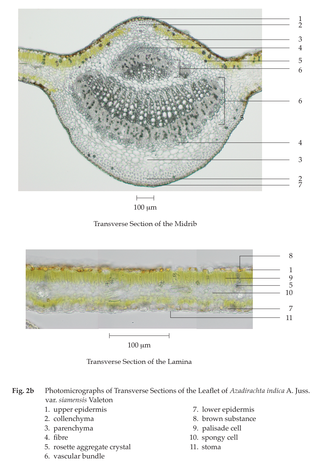
Microscopical (Figs. 2a, 2b, 2c) Transverse section of the leaflet through midrib and lamina shows upper epidermis, mesophyll, vascular bundle, and lower epidermis. Upper epidermis: 1 to 2 layers of slightly thick-walled rectangular cells, idioblasts, some containing brownish yellow substance, rosette aggregate crystals, covered with cuticle layer. Mesophyll: palisade cells, 1 to 3 layers, cylindrical, some containing rosette aggregate crystals; spongy cells, irregularly shaped, loosely arranged, varied in size, some containing rosette aggregate crystals; angular collenchyma and parenchyma, some containing rosette aggregate crystals or starch grains, in the upper and lower parts of the midrib. Vascular bundle: xylem and phloem, found in midrib and mesophyll. Lower epidermis: 1 to 2 layers of rectangular cells, some containing rosette aggregate crystals, slightly raised stomata, and glandular trichomes, covered with cuticle layer
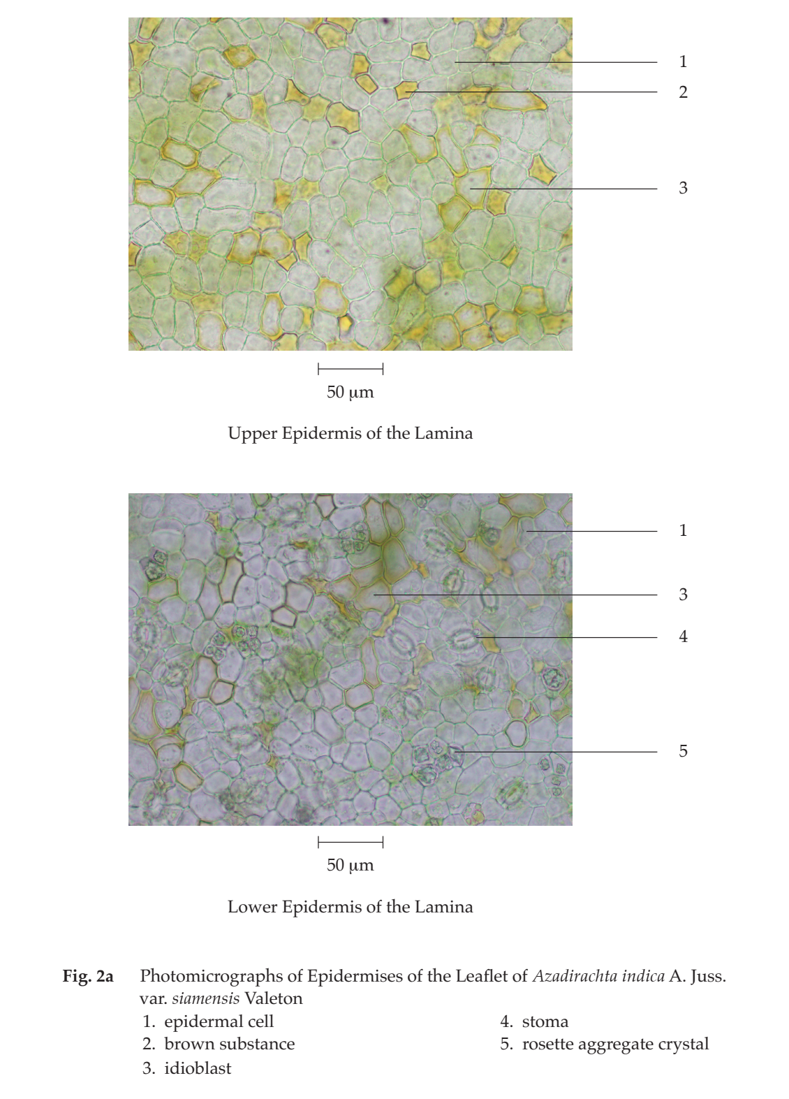
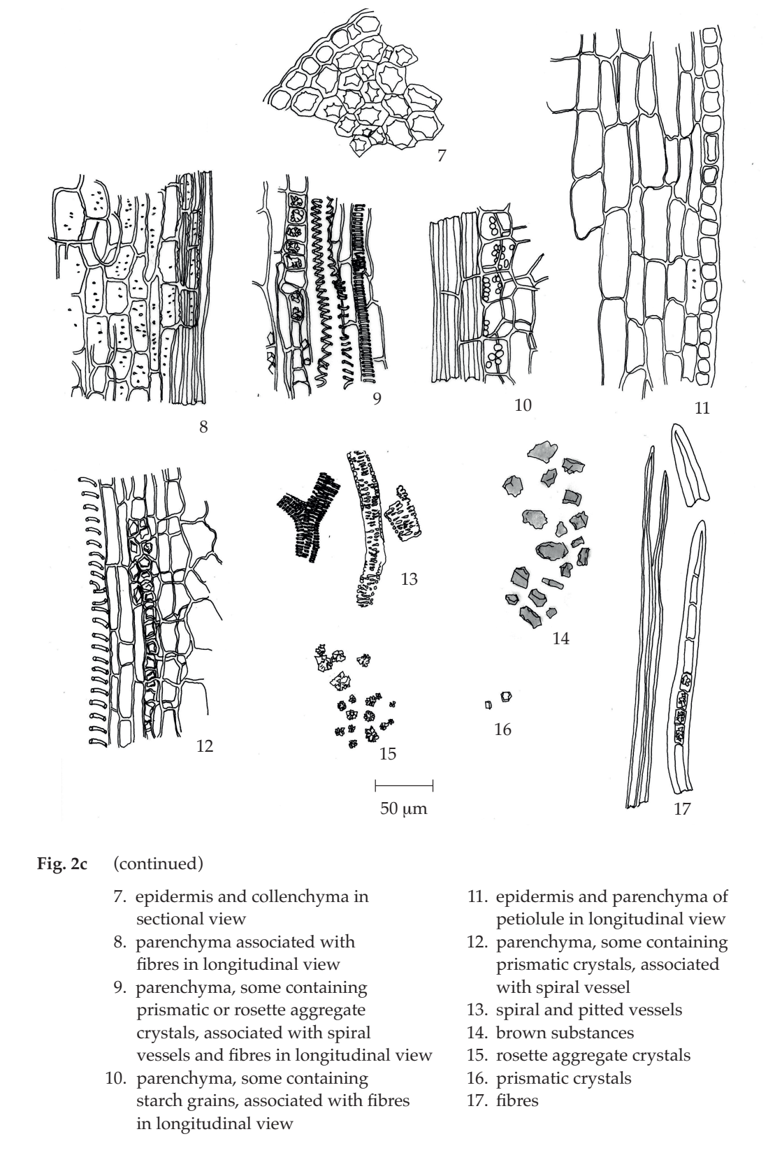
In surface view, upper and lower epidermises of the lamina show polygonal cells, some containing brown substance, idioblasts; stomata with 5 to 7 surrounding cells, some containing rosette aggregate crystals and glandular trichomes in the lower epidermis.
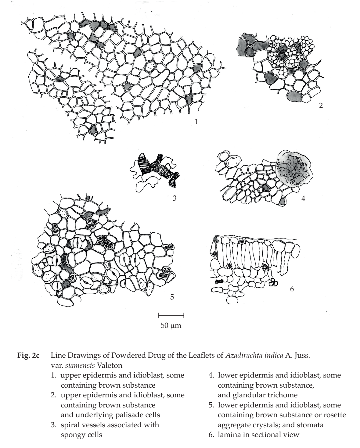
Siamese Neem Leaf in powder possesses the diagnostic microscopical characters of the unground drug. Both upper and lower epidermises with idioblast, some containing rosette aggregate crystals, and glandular trichome are characteristic.
Packaging and storage Siamese Neem Leaf shall be kept in well-closed containers, protected from light, and stored in a dry place.
Identification
A. Sonicate 5 g of the sample, in powder, with 30 mL of acetone for 30 minutes. Filter, discard the filtrate, and keep the marc. Extract the marc with the same solvent and procedure twice more. Further extract the marc with a mixture of 1 volume of a 1 per cent v/v solution of sulfuric acid and 100 volumes of a 40 per cent v/v solution of absolute ethanol by sonicating for 30 minutes and filter. Repeat the marc extraction two more times. Combine the resulting extracts (solution 1). To 1 mL of solution 1, add 1 piece of magnesium ribbon, shake well, and mix with 2 drops of hydrochloric acid: a faint pink colour develops.
B. To 200 mg of the sample, in powder, add 10 mL of water, heat in a water-bath for 10 minutes, and filter. To 2 mL of the filtrate, add a few drops of iron(III) chloride TS: a greenish blue colour is produced.
C. To 2.5 g of the sample, in powder, add 20 mL of ethanol, boil in a water-bath for 10 minutes and filter. To the filtrate, add 1 g of decolorizing charcoal, stir, and filter. Evaporate 3 mL of the filtrate to dryness, add 2 drops of a 2 per cent w/v solution of 3,5-dinitrobenzoic acid in methanol and 2 drops of a 5.7 per cent w/v solution of potassium hydroxide in methanol: a purplish red colour develops.
D. To 500 mg of the sample, in powder, add 10 mL of water, heat in a water-bath for 10 minutes and filter. To 2 mL of the filtrate, add a few drops of a 1 per cent w/v solution of gelatin in a 10 per cent w/v solution of sodium chloride: a white precipitate is produced.
E. Shake vigorously 500 mg of the sample, in powder, with 10 mL of water: a long lasting foam is produced.
F. Carry out the test as described in the “Thin-Layer Chromatography” (Appendix 3.1), using silica gel GF254 as the coating substance and a mixture of 40 volumes of ethyl acetate, 20 volumes of dichloromethane, 20 volumes of methanol, and 2 volumes of formic acid as the mobile phase and allowing the solvent front to ascend 8 cm above the line of application. Apply separately to the plate as bands of 8 mm, 10 µL of solution (A) and 5 µL of solution (B). For solution (A), use solution 1 as described in Identification A. For solution (B), dissolve 300 µg of rutin in 1 mL of methanol. After removal of the plate, allow it to dry in air, and examine the plate under ultraviolet light (254 nm), marking the quenching bands. The chromatogram obtained from solution (A) shows a quenching band (hRf value 32 to 33), corresponding to the rutin band from solution (B). Examine the plate under ultraviolet light (366 nm); the band due to rutin is blue and other six blue fluorescent bands are also observed. Heat the plate at 80º for at least 10 minutes and spray the plate with natural products (NP) TS while the plate is still warm. Subsequently spray the plate with polyethyleneglycol (PEG) TS and observe the colours of the bands under ultraviolet light (366 nm) within 5 to 15 minutes; the band due to rutin is orange and other four orange bands are also observed. Examine the plate under ultraviolet light (366 nm); the band due to rutin shows orange fluorescence. Other four orange and four blue fluorescent bands are also observed (Fig. 3)
Loss on drying Not more than 11.0 per cent w/w after drying at 105° to constant weight (Appendix 4.15).
Foreign matter Not more than 2.0 per cent w/w (Appendix 7.2).
Acid-insoluble ash Not more than 1.0 per cent w/w (Appendix 7.6).
Total ash Not more than 12.0 per cent w/w (Appendix 7.7).
Ethanol-soluble extractive Not less than 5.0 per cent w/w (Appendix 7.12).
Water-soluble extractive Not less than 20.0 per cent w/w (Appendix 7.12).
