ตำรามาตรฐานยาสมุนไพรไทย
Thai Herbal Pharmacopoeia
สำนักยาและวัตถุเสพติด กรมวิทยาศาสตร์การแพทย์ กระทรวงสาธารณสุข
Bureau of Drug and Narcotic, Department of Medical Sciences, Ministry of Public Healthสำนักยาและวัตถุเสพติด กรมวิทยาศาสตร์การแพทย์ กระทรวงสาธารณสุข
Bureau of Drug and Narcotic, Department of Medical Sciences, Ministry of Public Health(Tinospora crispa (L.) Hook.f. & Thomson)
(Nelumbo nucifera Gaertn.)
(Centella asiatica (L.) Urb.)
(Centella Dry Extract)
(Centella Cream)
(Mesua ferrea L.)
(Piper sarmentosum Roxb.)
(Piper sarmentosum Roxb.)
(Pterocarpus santalinus L. f.)
(Santalum album L.)
(Senna tora (L.) Roxb.)
(Senna alata (L.) Roxb.)
(Senna Alata Tea)
(Piper retrofractum Vahl)
(Myristica fragrans Houtt)
(Andrographis paniculata (Burm. f.) Nees)
(Andrographis Capsules)
(Allium ascalonicum L.)
(Ocimum tenuiflorum L.)
(Curcuma longa L.)
(Turmeric Capsules)
(Turmeric Dry Extract)
(Turmeric Dry Extract Capsules)
(Arcangelisia flava (L.) Merr.)
(Curcuma sp.)
Harrisonia perforata (Blanco) Merr.
(Aristolochia pierrei Lecomte)
(Zingiber officinale Roscoe)
(Ginger Capsules)
(Ginger Tea)
(Cassia fistula L.)
(Nardostachys jatamansi (D. Don) DC.)
(Angelica sinensis (Oliv.) Diels)
Artemisia annua L.
(Ligusticum sinense Oliv. cv. Chuanxiong)
(Neopicrorhiza scrophulariiflora Pennell)
(Atractylodes lancea (Thunb.) DC.)
(Aucklandia lappa Decne)
(Terminalia chebula Retz.)
(Angelica dahurica (Hoffm.) Benth. & Hook. f. ex Franch. & Sav. var. dahurica)
(Kaempferia parviflora Wall. ex Baker)
(Hibiscus sabdariffa L.)
(Roselle Tea)
(Allium sativum L.)
(Zingiber zerumbet (L.) Sm.)
(Wurfbainia testacea (Ridl.) Škorničk.& A. D. Poulsen)
(Cannabis sativa L.)
(Myristica fragrans Houtt)
(Dracaena cochinchinensis (Lour.) S. C. Chen)
(Ficus racemosa L.)
(Hyptis suaveolens (L.) Poit.)
Clerodendrum indicum (L.) Kuntze
(Phyllanthus emblica L.)
(Citrus hystrix DC.)
(Citrus hystrix DC.)
(Areca catechu L.)
(Momordica charantia L.)
Moringa oleifera Lam.
(Aegle marmelos (L.) Corrêa)
(Solanum trilobatum L.)
(Morus alba L.)
Gynostemma pentaphyllum(Thunb.)
Makino
(Clinacanthus nutans (Burm. f.) Lindau)
(Cissus quadrangularis L.)
(Mimusops elengi L.)
(Zingiber montanum (J. König) Link. ex A. Dietr.)
(Piper betle L.)
(Capsicum annuum L.)
(Capsicum Oleoresin)
(Capsicum Gel)
(Piper nigrum L.)
(Piper nigrum L.)
(Eurycoma longifolia Jack)
(Thunbergia laurifolia Lindl.)
(Piper wallichii (Miq.) Hand.-Mazz.)
Senna garrettiana (Craib) H. S. Irwin & Barneby
(Terminalia bellirica (Gaertn.) Roxb.)
(Terminalia chebula Retz.)
(Caesalpinia bonduc (L.) H. Roxb.)
(Tarlmounia elliptica (DC.) H. Rob., S. C. Keeley, Skvaria & R. Chan)
(Hog Creeper Vine Dry Extract Capsiles)
(Hog Creeper Vine Dry Extract)
(Brachypterum scandens (Roxb.) Miq.)
(Lepidium sativum L.)
(Nigella sativa L.)
(Cuminum cyminum L.)
(Foeniculum vulgare Mill.)
(Plantago ovata Forssk.)
(Pimpinella anisum L.)
(Carum carvi L.)
(Anethum graveolens L.)
(Trachyspermum ammi (L.) Sprague)
Albizia procera (Roxb.) Benth.
(Acorus calamus L.)
(Tiliacora triandra (Colebr.) Diels)
Cyanthillium cinereum (L.) H. Rob.
(Orthosiphon aristatus (Blume) Miq.)
Murdannia loriformis (Hassk.) R. S. Rao & Kammathy
(Capparis micracantha DC.)
(Chrysopogon zizanioides (L.) Roberty)
(Cyperus rotundus L.)
(Cannabis sativa L.)
(Syzygium aromaticum (L.) Merr. & L. M. Perry)
(Boesenbergia rotunda (L.) Mansf.)
(Acanthus ebracteatus Vahl)
(Acanthus ilicifolius L.)
(Kaempferia galanga L.)
(Curcuma comosa Roxb.)
Betula alnoides Buch.-Ham. ex D. Don
Cannabis sativa L.
Carthamus tinctorius L
Mitragyna speciosa (Korth.) Havil
Mallotus repandus (Rottler) Müll. Arg
Azadirachta indica A. Juss. var. siamensis Valeton
Azadirachta indica A. Juss. var. siamensis Valeton
Punica granatum L.
Rhinacanthus nasutus (L.) Kurz
Baliospermum solanifolium (Burm.) Suresh
Curcuma aeruginosa Roxb
Boesenbergia kingii Mood & L. M. Prince
Senegalia rugata (Lam.) Britton & Rose
Acacia concinna (Willd.) DC.
Senegalia rugata (Lam.) Britton & Rose
Acacia concinna (Willd.) DC.
Senna alexandriana Mill. var. alexandriana
Cassia acutifolia Delile, Cassia angustifolia Vahl
Butea superba Roxb. ex Willd.
[Plaso superba (Roxb. ex Willd.) Kuntze, Rudolphia superba (Roxb. ex Willd.) Poir.
Pueraria candollei Graham
ex Benth. var. mirifica (Airy Shaw & Suvat.) Niyomdham
Streblus asper Lour.
Suregada multiflora (A. Juss.) Baill. (Gelonium
multiflorum A. Juss.
Plumbago zeylanica L.
Plumbago indica L.
Biancaea sappan (L.) Tod.
Ziziphus attopensis Pierre
Streblus asper Lour.
Justicia gendarussa Burm. f.
Enhalus acoroides (L. f.) Royle
Bridelia ovata Decne.
Tamarindus indica L.
Citrus × aurantiifolia (Christm.) Swingle
Garcinia mangostana L.
Blumea balsamifera (L.) DC
Persicaria odorata (Lour.) Soják
Zingiber montanum (J. König) Link ex A. Dietr.
Mammea siamensis (Miq.) T. Anderson
Citrus maxima (Burm.) Merr.
Citrus × aurantium L. ‘Som Sa'
Punica granatum L.
Rhinacanthus nasutus (L.) Kurz
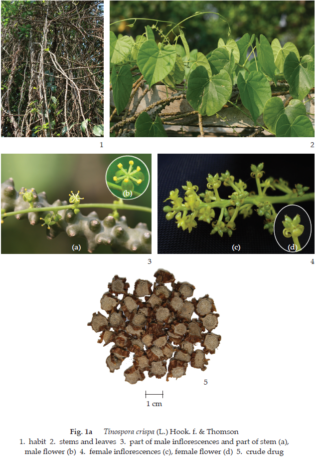
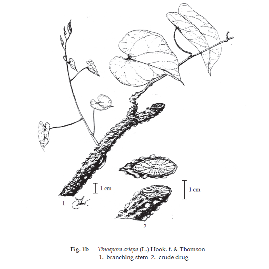
Tinospora Crispa Stem is the dried stem of Tinospora crispa (L.) Hook. f. & Thomson [T. crispa Diels, T. rumphii Boerl., T. tuberculata (Lam.) Beumée ex K. Heyne, T. gibbericaulis Hand. Mazz., T. mastersii Diels, T. thorelii Gagnep.] (Family Menispermaceae), Herbarium Specimen Number: DMSC 354, 355.
Constituents Tinospora Crispa Stem contains tinosporine, tinosporidine, picroretin, N-trans-feruloyl tyramine, N-cis-feruloyl tyramine, tinotuberide, borapetoside A, borapetol A, ceryl alcohol, β-sitosterol, stigmasterol, etc.
Description of the plant (Figs. 1a, 1b) Woody climber with tuberous roots; young stems smooth, older ones very prominently tuberculate with exceedingly bitter sap; aerial root filiform, very long. Leaves broadly ovate to orbicular, 5 to 14 cm long, 4 to 12 cm wide, apex acuminate, base cordate, palmately 5- to 7-nerved at the base; petiole 5 to 15 cm long. Inflorescence pseudoracemose, not coetaneous with the leaves. Male inflorescence very slender, a few in groups. Male flower small, on filiform pedicel; sepals pale green, 3 outer ones ovate, 3 inner ones obovate; petals 3; stamens 6. Female inflorescence similar to male onebut shorter. Female flower with sepals and petals as in male; s taminodes 6; carpels 3. Drupe orange, ellipsoid, up to 2 cm long.
Description Odour, indistinct; taste, intensely bitter.
Macroscopical (Fig. 1a) Cylindrical, transverse or oblique pieces, 3 to 30 mm long, 3 to 18 mm in diameter; externally brown, longitudinally wrinkled and numerous warty lenticels; internally pale greyish yellow, exhibiting a bark from 1.5 to 2.5 mm in thickness; surface, radiate and a minute disintegrated pith.
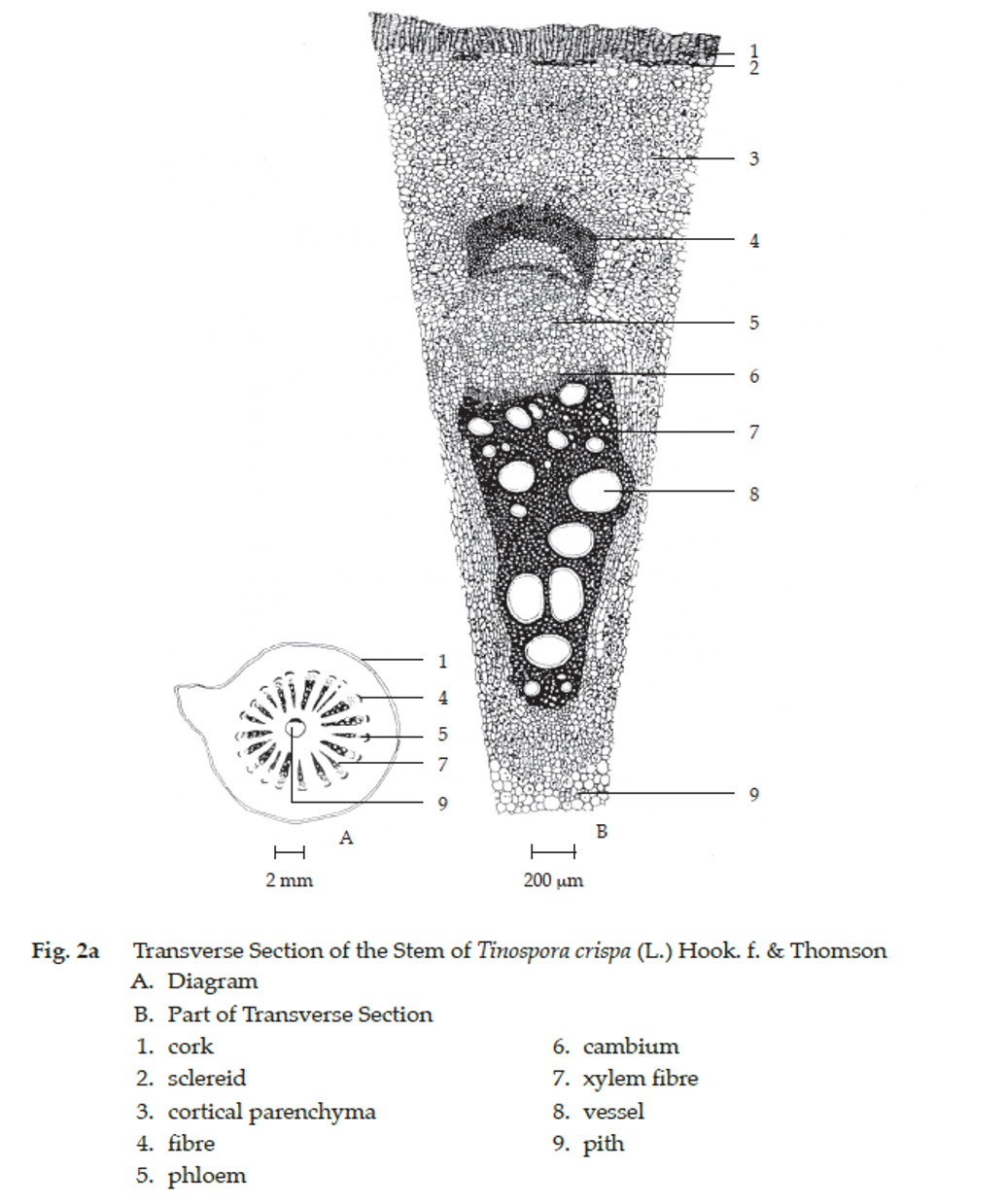
Microscopical (Figs. 2a, 2b) Transverse and longitudinal sections of the stem show cork, several layers of rectangular brownish cells. Cortex, broad zone of parenchyma cells containing starch granules, groups of stone cells (sclereids) containing prismatic crystals, occur beneath cork layers; parenchyma cells containing prismatic crystals occur in the innermost part of cortex adjacent to bast fibres. Stele composed of phloem and xylem separated by cambium, occurring several bands from cortex to pith, with medullary ray between the bands. Phloem composed of thick-walled bas t fibres and phloem tissue;
cambium, several layers of rectangular cells; xylem composed of large size vessels, xylem fibres and xylem parenchyma containing prismatic crystals; annular, spiral, reticulate, pitted and bordered-pitted vessels, up to 160 μm in diameter; medullary rays, non-lignified parenchyma containing s tarch granules. Pith, parenchyma cells containing starch granules.
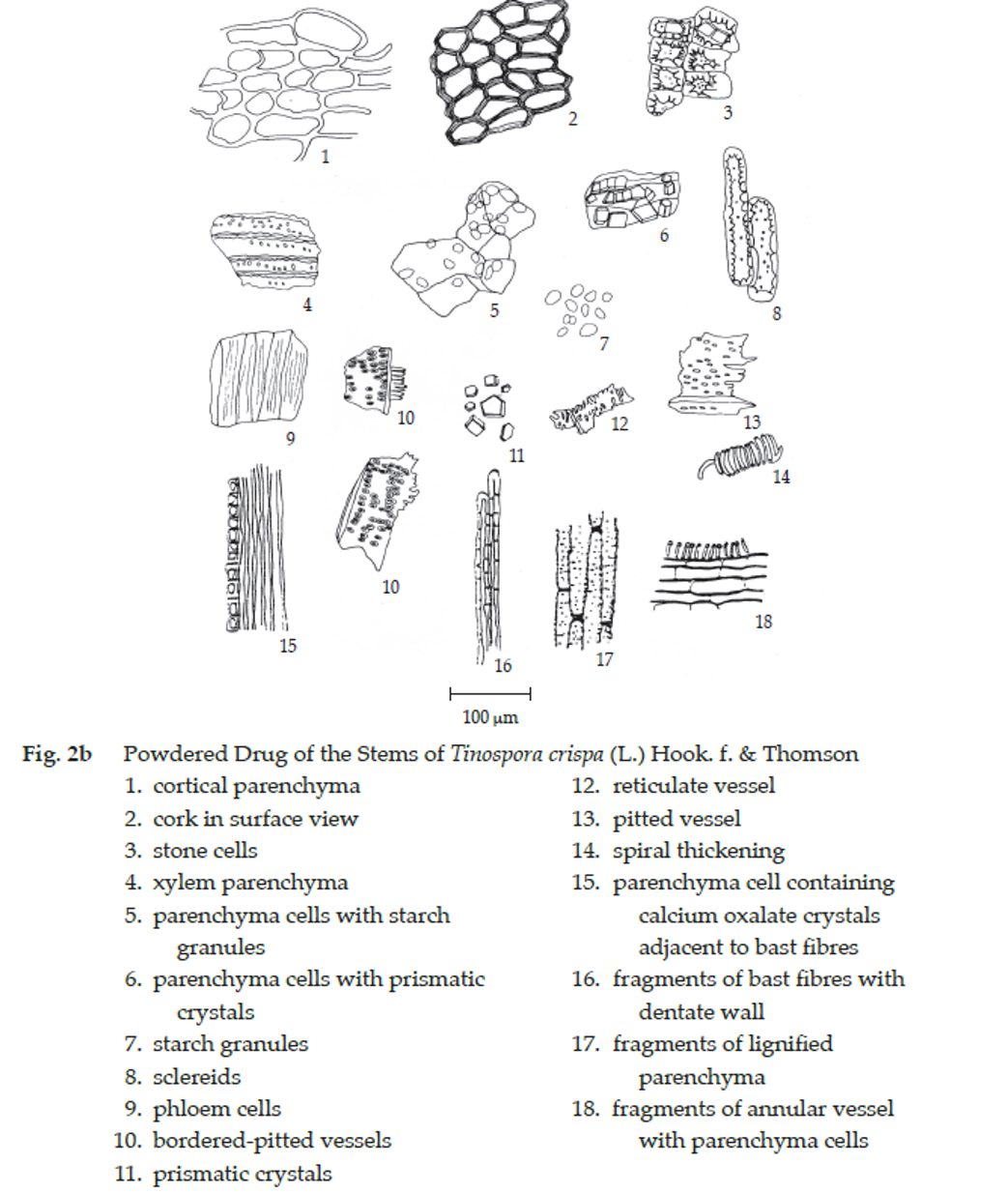
Tinospora Crispa Stem in powder possesses the diagnostic microscopical characters of the unground drug.
Packaging and storage Tinospora Crispa Stem shall be kept in well-closed containers, protected from light, and stored in a dry place.
Identification
A. To 500 mg of the sample, in powder, add 2 mL of acetic anhydride, warm on a water-bath for 2 minutes and filter. Slowly add 1 mL of sulfuric acid to the filtrate to form a layer: a brownish red ring forms at the zone of contact.
B. Shake vigorously 200 mg of the sample, in powder, with 10 mL of water: a long lasting foam is produced.
C. Add 10 mL of methanol to 1 g of the sample, in powder, warm on a water-bath for 10 minutes, shake intermittently, cool, and filter. To 1 mL of the filtrate, add a few drops of acetic potassium iodobismuthate TS: a brown precipitate forms.
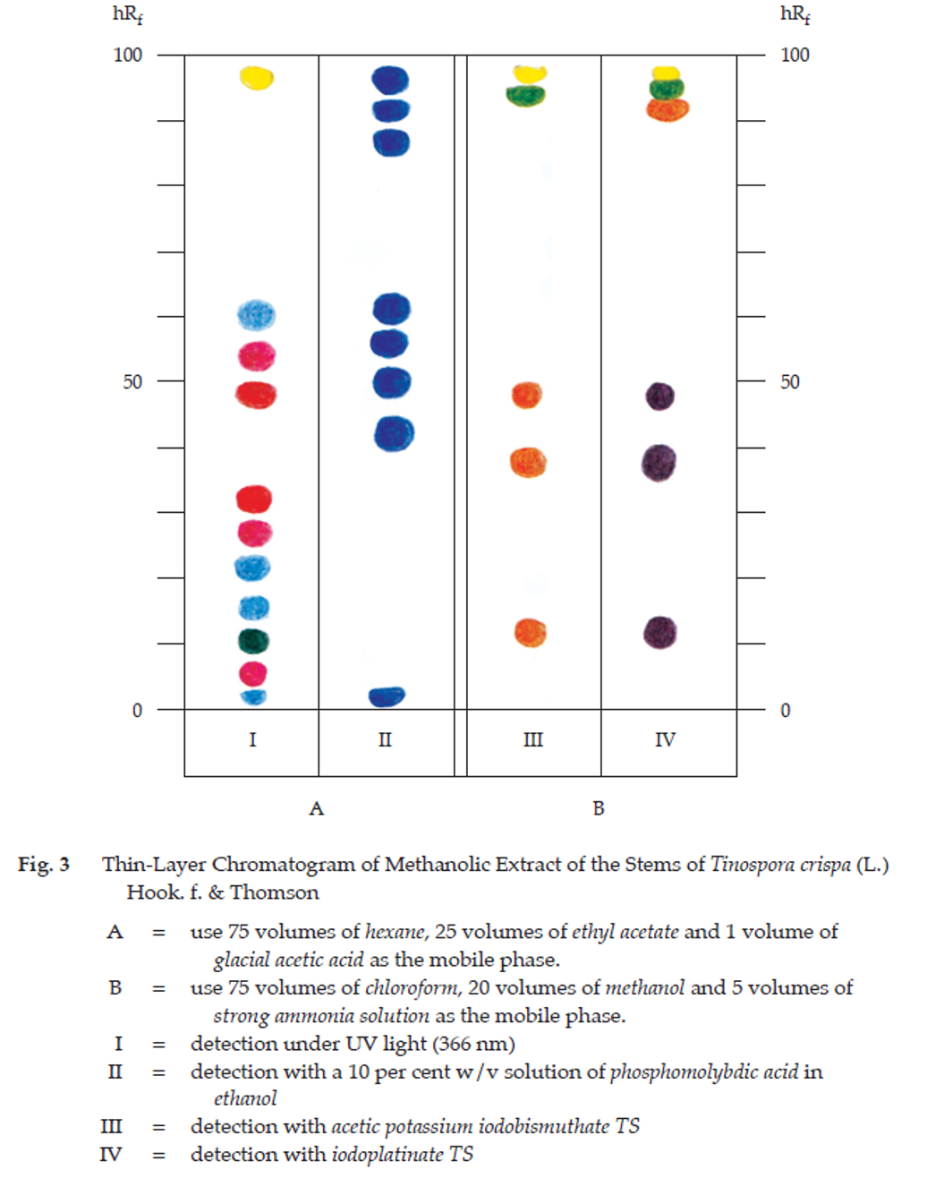
D. Test a Carry out the test as described in the “Thin-Layer Chromatography” (Appendix 3.1), using silica gel G as the coating substance and a mixture of 75 volumes of hexane, 25 volumes of ethyl acetate and 1 volume of glacial acetic acid as the mobile phase and allowing the solvent front to ascend 12 cm above the line of application. Apply to the plate, 20 μL of the test solution prepared by warming 1 g of the sample, in powder, with 10 mL of methanol on a water-bath at 60° for 10 minutes, shaking, filtering, and evaporating to 2 mL. After removal of the plate, allow it to dry in air and examine under ultraviolet light (366 nm), marking the spots. Several spots of different colours are observed (Table 1); see also Fig. 3. Spray the plate with a 10 per cent w/v solution of phosphomolybdic acid in ethanol, and heat at 100° for 5 to 10 minutes. Several blue spots are observed (Table 1); see also Fig. 3.
Test b Carry out the test as described in the “Thin-Layer Chromatography” (Appendix 3.1), using silica gel G as the coating substance and a mixture of 75 volumes of chloroform, 20 volumes of methanol and 5 volumes of strong ammonia solution as the mobile phase and allowing the solvent front to ascend 12 cm above the line of application. Apply to the plate, 20 μL of the test solution prepared as described in Test a. After removal of the plate, allow it to dry in air and spray with acetic potassium iodobismuthate TS. Several spots of different colours are observed (Table 1); see also Fig. 3. Repeat the same procedure on another plate but spray with iodoplatinate TS. Several spots of different colours are observed (Table 1); see also Fig. 3.
Table 1 hRf Values of Components in Methanol Extract of the Stems of Tinospora crispa (L.) Hook. f. & Thomson
| Spot | hRf Value | Detection | |||
| Mobile Phase I | Mobile Phase II | ||||
| UV 366 | 10 Per Cent W/V Solution of Phosphomolybdic Acid in Ethanol | Acetic Potassium Iodobismuthate TS | Iodoplatinate TS | ||
| 1 | 1-3 | light blue | dark blue | - | - |
| 2 | 4-7 | carmine red | - | - | - |
| 3 | 9-12 | moss green | - | - | - |
| 4 | 12-17 | light blue | - | - | - |
| 5 | 14-16 | - | - | orange | purple |
| 6 | 20-24 | light blue | - | - | - |
| 7 | 25-29 | carmine red | - | - | - |
| 8 | 31-34 | red | - | - | - |
| 9 | 37-40 | - | - | orange | purple |
| 10 | 42-45 | - | dark blue | - | - |
| 11 | 45-50 | red | - | - | - |
| 12 | 49-51 | - | - | orange | purple |
| 13 | 51-55 | carmine red | dark blue | - | - |
| 14 | 55-58 | - | dark blue | - | - |
| 15 | 59-63 | light blue | - | - | - |
| 16 | 61-64 | - | dark blue | - | - |
| 17 | 86-90 | - | dark blue | - | - |
| 18 | 91-94 | - | dark blue | - | - |
| 19 | 92-96 | - | - | - | orange |
| 20 | 96-98 | yellow | dark blue | moss green | moss green |
| 21 | 98-99 | - | - | yellow | yellow |
Mobile Phase I: 75 volumes of hexane, 25 volumes of ethyl acetate and 1 volume of glacial acetic acid
Mobile Phase II: 75 volumes of chloroform, 20 volumes of methanol and 5 volumes of strong ammonia solution
Loss on drying Not more than 11.0 per cent w/w after drying 10 g at 105° for 5 hours (Appendix 4.15).
Foreign matter Not more than 2.0 per cent w/w (Appendix 7.2).
Acid-insoluble ash Not more than 0.5 per cent w/w (Appendix 7.6).
Total ash Not more than 7.0 per cent w/w (Appendix 7.7).
Ethanol-soluble extractive Not less than 5.0 per cent w/w (Appendix 7.12).
Water-soluble extractive Not less than 10.0 per cent w/w (Appendix 7.12).
Determination of bitterness Not less than 210 units per g when determined by the following method.
Standard preparation Transfer about 100 mg of quinine hydrochloride, accurately weighed, to a 100-mL volumetric flask, dissolve in safe drinking water, dilute to volume with the same solvent, and mix. Dilute this solution quantitatively with safe drinking water to obtain the solution containing 10 µg of quinine hydrochloride per mL. This solution is used as the stock solution of quinine hydrochloride (SQ).
Prepare a serial dilution of SQ in nine test-tubes according to the following table for the first series of testing.
| No. of Tubes | 1 | 2 | 3 | 4 | 5 | 6 | 7 | 8 | 9 |
| mL of SQ | 4.2 | 4.4 | 4.6 | 4.8 | 5.0 | 5.2 | 5.4 | 5.6 | 5.8 |
| mL of water | 5.8 | 5.6 | 5.4 | 5.2 | 5.0 | 4.8 | 4.6 | 4.4 | 4.2 |
| µg of quinine hydrochloride | 42 | 44 | 46 | 48 | 50 | 52 | 54 | 56 | 58 |
Test preparation Transfer about 200 mg of the sample, in powder, accurately weighed, into a 100-mL conical flask, add 45 mL of safe drinking water, and reflux in a boiling water-bath for 1 hour with frequent shaking. Cool, filter and dilute the filtrate with safe drinking water to 50.0 mL. Pipette 1.0 mL of this solution into a 100-mL volumetric flask and dilute with safe drinking water to volume. This solution is used as the stock solution of the sample (ST). Calculate its concentration and express it in µg per mL.
Prepare a serial dilution of ST in 10 test-tubes according to the following table for the second series of testing.
| No. of Tubes | 1 | 2 | 3 | 4 | 5 | 6 | 7 | 8 | 9 | 10 |
| mL of ST (b) | 1.0 | 2.0 | 3.0 | 4.0 | 5.0 | 6.0 | 7.0 | 8.0 | 9.0 | 10.0 |
| mL of water | 9.0 | 8.0 | 7.0 | 6.0 | 5.0 | 4.0 | 3.0 | 2.0 | 1.0 | - |
Procedure After rinsing the mouth with safe drinking water, taste 10 mL of the dilution swirling it in the mouth mainly near the base of the tongue for 30 seconds. Unless otherwise specified, always begin with the lowest concentration of the serial dilution. If the bitter sensation is no longer felt, withdraw the solution and wait for 1 minute to ascertain that there is no delayed sensitivity. Then rinse with safe drinking water. The next highest concentration of dilution should not be tasted until at least 10 minutes have passed. The threshold bitter concentration is the lowest concentration of dilution at which a material still provokes a bitter sensation. After the first series of tests, rinse the mouth thoroughly with safe drinking water, until no bitter sensation remains and wait for at least 10 minutes before carrying out the second series of tests. In this series of testing and in order to save time, it is advisable to first ascertain whether the solution in tube no. 5 (containing 5 mL of ST in 10 mL) gives a bitter sensation. If noted, find the threshold bitter concentration of the material by tasting the dilutions in tubes nos. 1 to 4. If the solution in tube no. 5 does not give a bitter sensation, find the threshold bitter concentration in the dilutions of tubes nos. 6 to 10. All solutions and safe drinking water for mouthwashing should be at 20° to 25°.