ตำรามาตรฐานยาสมุนไพรไทย
Thai Herbal Pharmacopoeia
สำนักยาและวัตถุเสพติด กรมวิทยาศาสตร์การแพทย์ กระทรวงสาธารณสุข
Bureau of Drug and Narcotic, Department of Medical Sciences, Ministry of Public Healthสำนักยาและวัตถุเสพติด กรมวิทยาศาสตร์การแพทย์ กระทรวงสาธารณสุข
Bureau of Drug and Narcotic, Department of Medical Sciences, Ministry of Public Health(Tinospora crispa (L.) Hook.f. & Thomson)
(Nelumbo nucifera Gaertn.)
(Centella asiatica (L.) Urb.)
(Centella Dry Extract)
(Centella Cream)
(Mesua ferrea L.)
(Piper sarmentosum Roxb.)
(Piper sarmentosum Roxb.)
(Pterocarpus santalinus L. f.)
(Santalum album L.)
(Senna tora (L.) Roxb.)
(Senna alata (L.) Roxb.)
(Senna Alata Tea)
(Piper retrofractum Vahl)
(Myristica fragrans Houtt)
(Andrographis paniculata (Burm. f.) Nees)
(Andrographis Capsules)
(Allium ascalonicum L.)
(Ocimum tenuiflorum L.)
(Curcuma longa L.)
(Turmeric Capsules)
(Turmeric Dry Extract)
(Turmeric Dry Extract Capsules)
(Arcangelisia flava (L.) Merr.)
(Curcuma sp.)
Harrisonia perforata (Blanco) Merr.
(Aristolochia pierrei Lecomte)
(Zingiber officinale Roscoe)
(Ginger Capsules)
(Ginger Tea)
(Cassia fistula L.)
(Nardostachys jatamansi (D. Don) DC.)
(Angelica sinensis (Oliv.) Diels)
Artemisia annua L.
(Ligusticum sinense Oliv. cv. Chuanxiong)
(Neopicrorhiza scrophulariiflora Pennell)
(Atractylodes lancea (Thunb.) DC.)
(Aucklandia lappa Decne)
(Terminalia chebula Retz.)
(Angelica dahurica (Hoffm.) Benth. & Hook. f. ex Franch. & Sav. var. dahurica)
(Kaempferia parviflora Wall. ex Baker)
(Hibiscus sabdariffa L.)
(Roselle Tea)
(Allium sativum L.)
(Zingiber zerumbet (L.) Sm.)
(Wurfbainia testacea (Ridl.) Škorničk.& A. D. Poulsen)
(Cannabis sativa L.)
(Myristica fragrans Houtt)
(Dracaena cochinchinensis (Lour.) S. C. Chen)
(Ficus racemosa L.)
(Hyptis suaveolens (L.) Poit.)
Clerodendrum indicum (L.) Kuntze
(Phyllanthus emblica L.)
(Citrus hystrix DC.)
(Citrus hystrix DC.)
(Areca catechu L.)
(Momordica charantia L.)
Moringa oleifera Lam.
(Aegle marmelos (L.) Corrêa)
(Solanum trilobatum L.)
(Morus alba L.)
Gynostemma pentaphyllum(Thunb.)
Makino
(Clinacanthus nutans (Burm. f.) Lindau)
(Cissus quadrangularis L.)
(Mimusops elengi L.)
(Zingiber montanum (J. König) Link. ex A. Dietr.)
(Piper betle L.)
(Capsicum annuum L.)
(Capsicum Oleoresin)
(Capsicum Gel)
(Piper nigrum L.)
(Piper nigrum L.)
(Eurycoma longifolia Jack)
(Thunbergia laurifolia Lindl.)
(Piper wallichii (Miq.) Hand.-Mazz.)
Senna garrettiana (Craib) H. S. Irwin & Barneby
(Terminalia bellirica (Gaertn.) Roxb.)
(Terminalia chebula Retz.)
(Caesalpinia bonduc (L.) H. Roxb.)
(Tarlmounia elliptica (DC.) H. Rob., S. C. Keeley, Skvaria & R. Chan)
(Hog Creeper Vine Dry Extract Capsiles)
(Hog Creeper Vine Dry Extract)
(Brachypterum scandens (Roxb.) Miq.)
(Lepidium sativum L.)
(Nigella sativa L.)
(Cuminum cyminum L.)
(Foeniculum vulgare Mill.)
(Plantago ovata Forssk.)
(Pimpinella anisum L.)
(Carum carvi L.)
(Anethum graveolens L.)
(Trachyspermum ammi (L.) Sprague)
Albizia procera (Roxb.) Benth.
(Acorus calamus L.)
(Tiliacora triandra (Colebr.) Diels)
Cyanthillium cinereum (L.) H. Rob.
(Orthosiphon aristatus (Blume) Miq.)
Murdannia loriformis (Hassk.) R. S. Rao & Kammathy
(Capparis micracantha DC.)
(Chrysopogon zizanioides (L.) Roberty)
(Cyperus rotundus L.)
(Cannabis sativa L.)
(Syzygium aromaticum (L.) Merr. & L. M. Perry)
(Boesenbergia rotunda (L.) Mansf.)
(Acanthus ebracteatus Vahl)
(Acanthus ilicifolius L.)
(Kaempferia galanga L.)
(Curcuma comosa Roxb.)
Betula alnoides Buch.-Ham. ex D. Don
Cannabis sativa L.
Carthamus tinctorius L
Mitragyna speciosa (Korth.) Havil
Mallotus repandus (Rottler) Müll. Arg
Azadirachta indica A. Juss. var. siamensis Valeton
Azadirachta indica A. Juss. var. siamensis Valeton
Punica granatum L.
Rhinacanthus nasutus (L.) Kurz
Baliospermum solanifolium (Burm.) Suresh
Curcuma aeruginosa Roxb
Boesenbergia kingii Mood & L. M. Prince
Senegalia rugata (Lam.) Britton & Rose
Acacia concinna (Willd.) DC.
Senegalia rugata (Lam.) Britton & Rose
Acacia concinna (Willd.) DC.
Senna alexandriana Mill. var. alexandriana
Cassia acutifolia Delile, Cassia angustifolia Vahl
Butea superba Roxb. ex Willd.
[Plaso superba (Roxb. ex Willd.) Kuntze, Rudolphia superba (Roxb. ex Willd.) Poir.
Pueraria candollei Graham
ex Benth. var. mirifica (Airy Shaw & Suvat.) Niyomdham
Streblus asper Lour.
Suregada multiflora (A. Juss.) Baill. (Gelonium
multiflorum A. Juss.
Plumbago zeylanica L.
Plumbago indica L.
Biancaea sappan (L.) Tod.
Ziziphus attopensis Pierre
Streblus asper Lour.
Justicia gendarussa Burm. f.
Enhalus acoroides (L. f.) Royle
Bridelia ovata Decne.
Tamarindus indica L.
Citrus × aurantiifolia (Christm.) Swingle
Garcinia mangostana L.
Blumea balsamifera (L.) DC
Persicaria odorata (Lour.) Soják
Zingiber montanum (J. König) Link ex A. Dietr.
Mammea siamensis (Miq.) T. Anderson
Citrus maxima (Burm.) Merr.
Citrus × aurantium L. ‘Som Sa'
Punica granatum L.
Rhinacanthus nasutus (L.) Kurz
Devil’s Backbone is the dried stem of Cissus quadrangularis L. (Family Vitaceae), Herbarium Specimen Number: DMSC 5158, Crude Drug Number: DMSc 0754.
Constituents Devil’s Backbone contains triterpenoids (e.g.,lupeol), flavonoids (e.g., kaempferol, quercetin), calcium oxalate, ascorbic acid, tannins, carotenoids, etc.
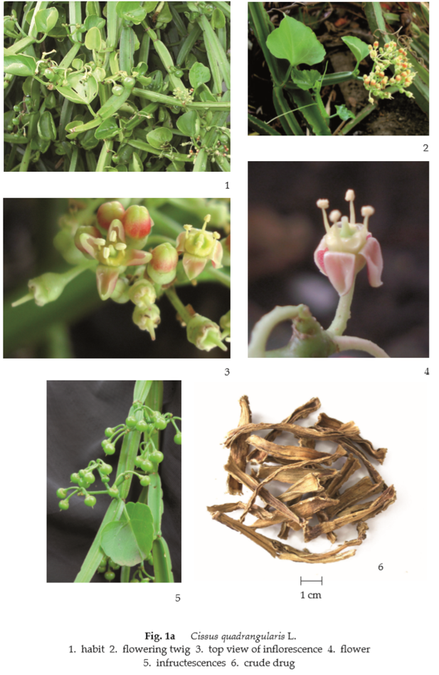
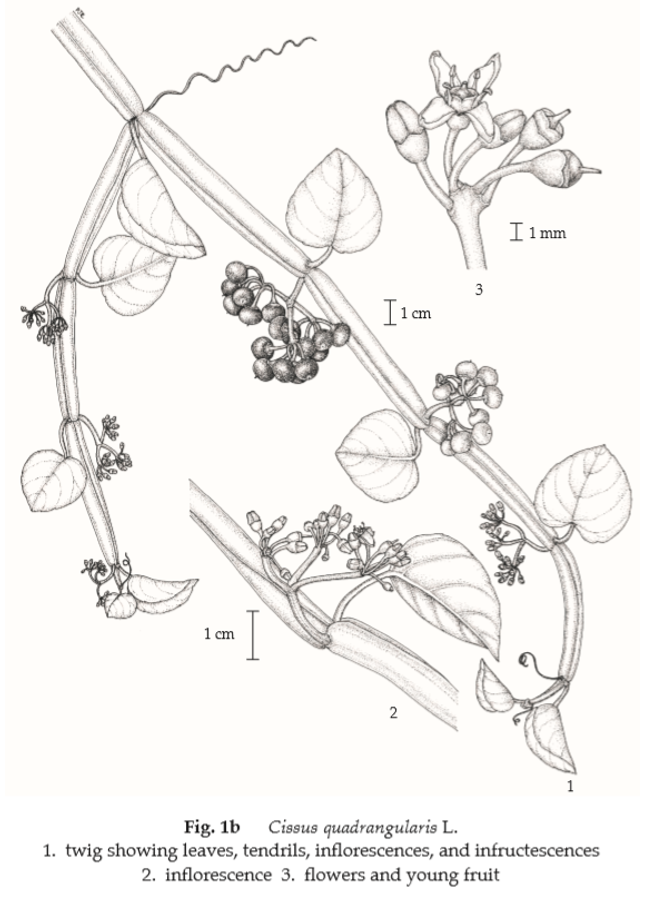
Description of the plant (Figs. 1a, 1b) Woody climber; stem green, succulent, quadrangular, angles winged, much constricted at nodes; tendril unequally bifurcate, 10 to 15 cm long, leaf-opposed; stipules 2, ovate, 3 to 4 mm long, 2 to 3 mm wide, caducous. Leaves simple, alternate, cordate to ovate, 4 to 7 cm long, 2.5 to 7 cm wide, apex obtuse to cuspidate, base auriculate to truncate, margin crenulate with minute teeth, coriaceous, with 3 main basal veins and 3 pairs of secondary veins, caducous; petiole up to 2 cm long, quadrangular. Inflorescence compound umbelliform cymes, leaf-opposed, up to 3 cm long, up to 5.5 cm wide; peduncle up to 2 cm long; pedicel up to 4 mm long. Flower small, reddish; calyx, cupuliform, inconspicuous lobes; petals 4, ovate, 2 to 3 mm long, 1 to 1.5 mm wide, apex hood-shaped; stamens 4, filament filiform, 0.7 to 1.3 mm long, anther elliptic-ovate; disc slightly 4-lobed; ovary superior, adnate to the disc, 2-loculed, ovules 2 per locule, style and stigma inconspicuously cylindrical. Fruit berry, mostly globose, 0.5 to 1 cm long, 0.5 to 1 cm wide, reddish purple to black. Seed(s) 1 or 2, orbicular or ellipsoid, about 5 mm long, about 4 mm wide, smooth.
Description Odour, mild; taste, bland.
Macroscopical (Fig. 1a) Part of quadrangular vine, varying in size and length, with or without node, pale brown colour; tendril occasionally found.
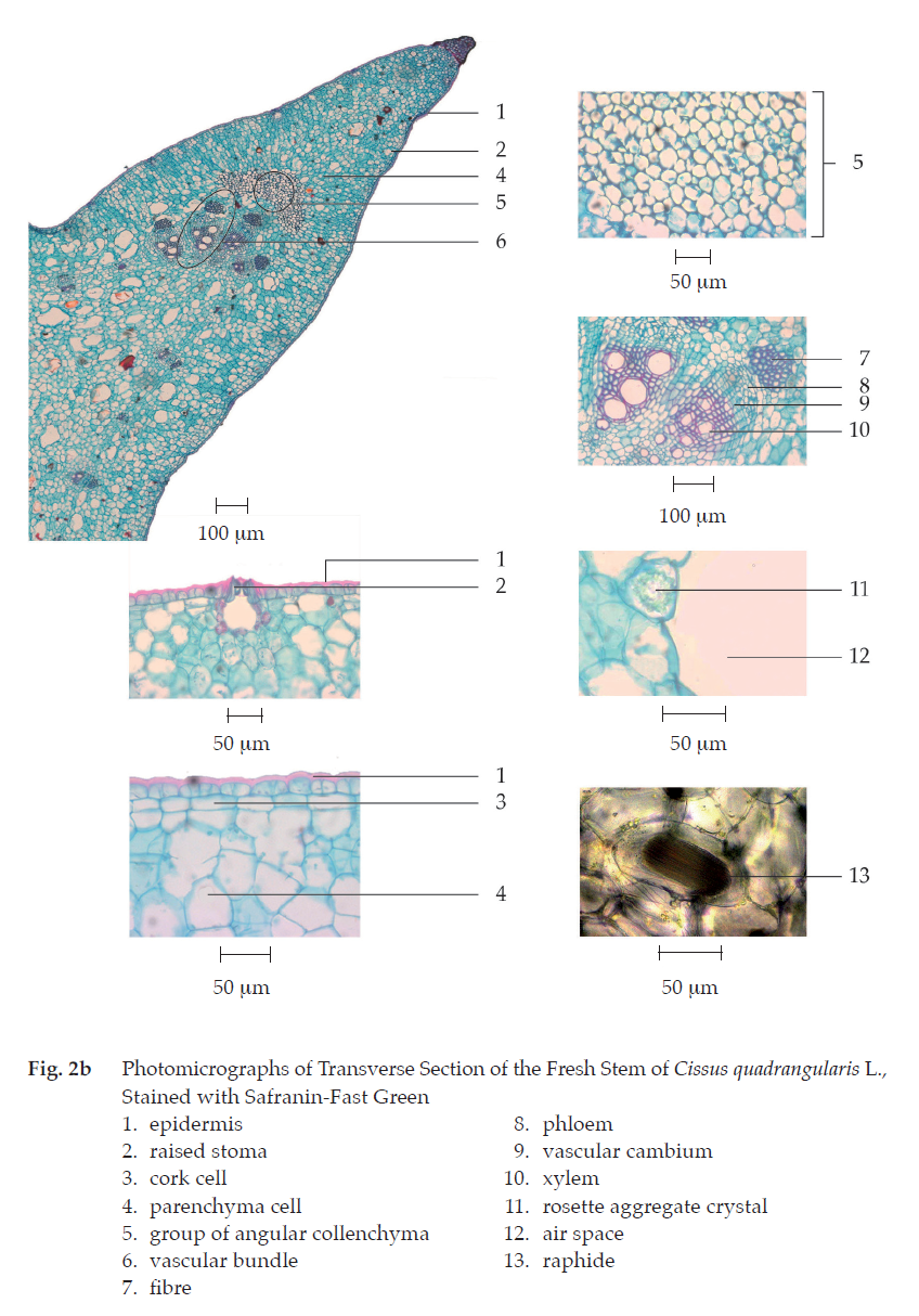
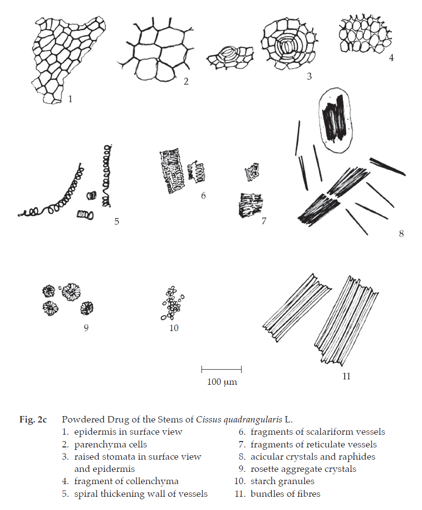
Microscopical (Figs. 2a, 2b, 2c) Transverse section of the fresh stem shows epidermal layer containing raised stomata, thick cuticle layer and underneath cork layer. Cortex layer composed of parenchyma, some of which contain starch granules, rosette aggregate and raphide crystals, a group of thick-walled angular collenchyma in each ridge and air spaces. Fibrovascular bundle consisting of fibres, phloem, vascular cambium, and xylem.
Devil’s Backbone in powder possesses the diagnostic microscopical characters of the unground drug.
Warning The fresh juice of the stem or the crushed-fresh stem irritates the skin and mucous membrane, and may cause itching.

Additional information
1. It is commonly used with other herbal drugs in the antihemorrhoidal remedies.
2. Traditionally, thin slices of one fresh internode wrapped with tamarind pulp or ripe banana are daily consumed for three consecutive days to treat hemorrhoid.
Packaging and storage Devil’s Backbone shall be kept in well-closed containers, protected from light, and stored in a dry place.
Identification
A. To 5 g of the sample, in powder, add 20 mL of ethanol, shake occasionally during 1 hour and filter (solution 1). To 2 mL of solution 1, add 4 or 5 pieces of magnesium ribbon, shake well and mix with a few drops of hydrochloric acid: a pink colour develops.
B. To 2 mL of solution 1, add a few drops of iron(III) chloride TS, and shake well: a blue colour develops.
C. Evaporate 2 mL of solution 1 to dryness, dissolve the residue in 2 mL of acetic anhydride, and then slowly add 1 mL of sulfuric acid to form two layers: a brown ring forms at the zone of contact and the upper layer is yellow which gradually changes to green.
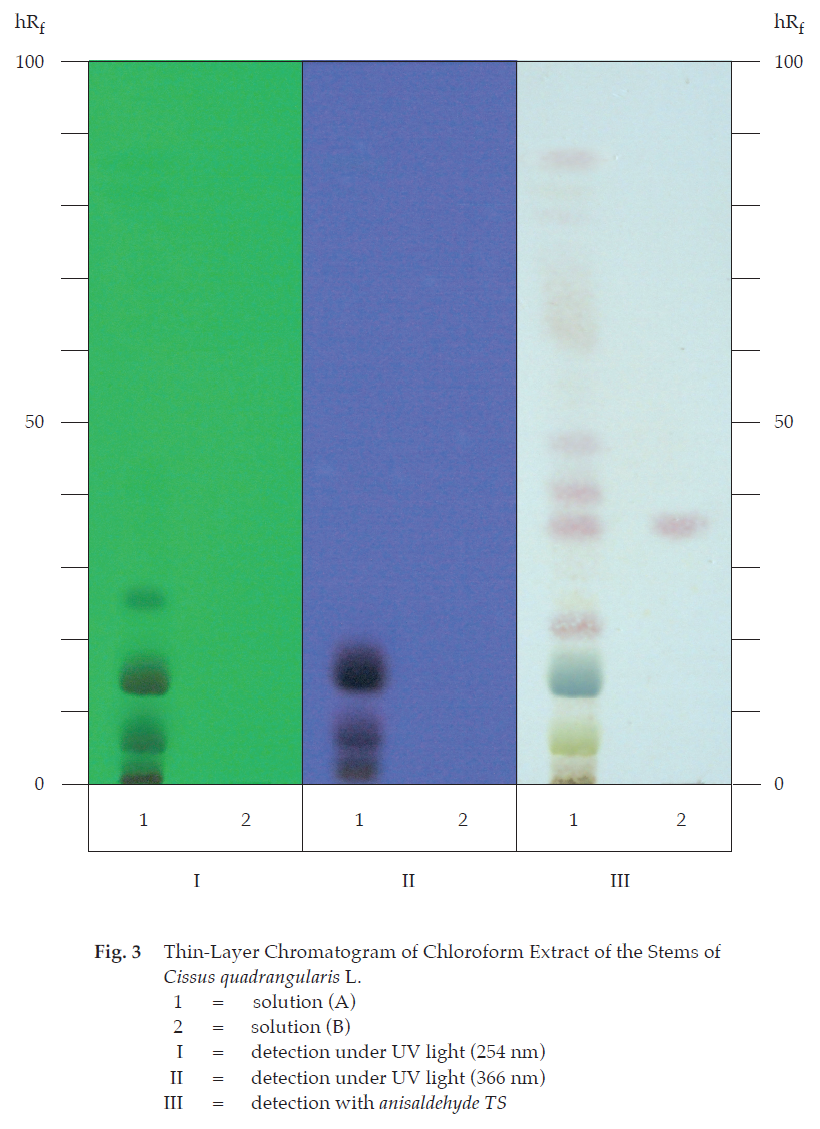
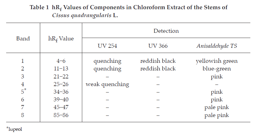
D. Carry out the test as described in the “Thin-Layer Chromatography” (Appendix 3.1), using silica gel GF254 as the coating substance and a mixture of 80 volumes of hexane and 10 volumes of ethyl acetate as the mobile phase and allowing the solvent front to ascend 12 cm above the line of application. Apply separately to the plate as bands of 5 mm, 5 μL each of the following two solutions. Prepare solution (A) by macerating 500 mg of the sample, in No. 150 powder, with 5 mL of chloroform, shaking frequently during 12 hours and filtering. Evaporate the filtrate to dryness and dissolve the residue in 1 ml of chloroform. For solution (B), dissolve 1 mg of lupeol in 1 mL of chloroform. After removal of the plate, allow it to dry in air and examine under ultraviolet light (254 nm), marking the quenching bands. Subsequently examine the plate under ultraviolet light (366 nm); two reddish black bands are observed. Spray the plate with anisaldehyde TS and heat at 110° until colours are fully developed; the chromatogram obtained from solution (A) shows a pink band (hRf value 34 to 36) corresponding to the lupeol band from solution (B). Several other bands of different colours are also observed (Table 1); see also Fig. 3.
Loss on drying Not more than 9.0 per cent w/w after drying at 105° for 5 hours (Appendix 4.15).
Foreign matter Not more than 5.0 per cent w/w (Appendix 7.2).
Acid-insoluble ash Not more than 1.5 per cent w/w (Appendix 7.6).
Total ash Not more than 20.0 per cent w/w (Appendix 7.7).
Ethanol (80 per cent)-soluble extractive Not less than 12.5 per cent w/w (Appendix 7.12).
Water-soluble extractive Not less than 25.0 per cent w/w (Appendix 7.12).
Dose 3 to 6 g a day.