ตำรามาตรฐานยาสมุนไพรไทย
Thai Herbal Pharmacopoeia
สำนักยาและวัตถุเสพติด กรมวิทยาศาสตร์การแพทย์ กระทรวงสาธารณสุข
Bureau of Drug and Narcotic, Department of Medical Sciences, Ministry of Public Healthสำนักยาและวัตถุเสพติด กรมวิทยาศาสตร์การแพทย์ กระทรวงสาธารณสุข
Bureau of Drug and Narcotic, Department of Medical Sciences, Ministry of Public Health(Tinospora crispa (L.) Hook.f. & Thomson)
(Nelumbo nucifera Gaertn.)
(Centella asiatica (L.) Urb.)
(Centella Dry Extract)
(Centella Cream)
(Mesua ferrea L.)
(Piper sarmentosum Roxb.)
(Piper sarmentosum Roxb.)
(Pterocarpus santalinus L. f.)
(Santalum album L.)
(Senna tora (L.) Roxb.)
(Senna alata (L.) Roxb.)
(Senna Alata Tea)
(Piper retrofractum Vahl)
(Myristica fragrans Houtt)
(Andrographis paniculata (Burm. f.) Nees)
(Andrographis Capsules)
(Allium ascalonicum L.)
(Ocimum tenuiflorum L.)
(Curcuma longa L.)
(Turmeric Capsules)
(Turmeric Dry Extract)
(Turmeric Dry Extract Capsules)
(Arcangelisia flava (L.) Merr.)
(Curcuma sp.)
Harrisonia perforata (Blanco) Merr.
(Aristolochia pierrei Lecomte)
(Zingiber officinale Roscoe)
(Ginger Capsules)
(Ginger Tea)
(Cassia fistula L.)
(Nardostachys jatamansi (D. Don) DC.)
(Angelica sinensis (Oliv.) Diels)
Artemisia annua L.
(Ligusticum sinense Oliv. cv. Chuanxiong)
(Neopicrorhiza scrophulariiflora Pennell)
(Atractylodes lancea (Thunb.) DC.)
(Aucklandia lappa Decne)
(Terminalia chebula Retz.)
(Angelica dahurica (Hoffm.) Benth. & Hook. f. ex Franch. & Sav. var. dahurica)
(Kaempferia parviflora Wall. ex Baker)
(Hibiscus sabdariffa L.)
(Roselle Tea)
(Allium sativum L.)
(Zingiber zerumbet (L.) Sm.)
(Wurfbainia testacea (Ridl.) Škorničk.& A. D. Poulsen)
(Cannabis sativa L.)
(Myristica fragrans Houtt)
(Dracaena cochinchinensis (Lour.) S. C. Chen)
(Ficus racemosa L.)
(Hyptis suaveolens (L.) Poit.)
Clerodendrum indicum (L.) Kuntze
(Phyllanthus emblica L.)
(Citrus hystrix DC.)
(Citrus hystrix DC.)
(Areca catechu L.)
(Momordica charantia L.)
Moringa oleifera Lam.
(Aegle marmelos (L.) Corrêa)
(Solanum trilobatum L.)
(Morus alba L.)
Gynostemma pentaphyllum(Thunb.)
Makino
(Clinacanthus nutans (Burm. f.) Lindau)
(Cissus quadrangularis L.)
(Mimusops elengi L.)
(Zingiber montanum (J. König) Link. ex A. Dietr.)
(Piper betle L.)
(Capsicum annuum L.)
(Capsicum Oleoresin)
(Capsicum Gel)
(Piper nigrum L.)
(Piper nigrum L.)
(Eurycoma longifolia Jack)
(Thunbergia laurifolia Lindl.)
(Piper wallichii (Miq.) Hand.-Mazz.)
Senna garrettiana (Craib) H. S. Irwin & Barneby
(Terminalia bellirica (Gaertn.) Roxb.)
(Terminalia chebula Retz.)
(Caesalpinia bonduc (L.) H. Roxb.)
(Tarlmounia elliptica (DC.) H. Rob., S. C. Keeley, Skvaria & R. Chan)
(Hog Creeper Vine Dry Extract Capsiles)
(Hog Creeper Vine Dry Extract)
(Brachypterum scandens (Roxb.) Miq.)
(Lepidium sativum L.)
(Nigella sativa L.)
(Cuminum cyminum L.)
(Foeniculum vulgare Mill.)
(Plantago ovata Forssk.)
(Pimpinella anisum L.)
(Carum carvi L.)
(Anethum graveolens L.)
(Trachyspermum ammi (L.) Sprague)
Albizia procera (Roxb.) Benth.
(Acorus calamus L.)
(Tiliacora triandra (Colebr.) Diels)
Cyanthillium cinereum (L.) H. Rob.
(Orthosiphon aristatus (Blume) Miq.)
Murdannia loriformis (Hassk.) R. S. Rao & Kammathy
(Capparis micracantha DC.)
(Chrysopogon zizanioides (L.) Roberty)
(Cyperus rotundus L.)
(Cannabis sativa L.)
(Syzygium aromaticum (L.) Merr. & L. M. Perry)
(Boesenbergia rotunda (L.) Mansf.)
(Acanthus ebracteatus Vahl)
(Acanthus ilicifolius L.)
(Kaempferia galanga L.)
(Curcuma comosa Roxb.)
Betula alnoides Buch.-Ham. ex D. Don
Cannabis sativa L.
Carthamus tinctorius L
Mitragyna speciosa (Korth.) Havil
Mallotus repandus (Rottler) Müll. Arg
Azadirachta indica A. Juss. var. siamensis Valeton
Azadirachta indica A. Juss. var. siamensis Valeton
Punica granatum L.
Rhinacanthus nasutus (L.) Kurz
Baliospermum solanifolium (Burm.) Suresh
Curcuma aeruginosa Roxb
Boesenbergia kingii Mood & L. M. Prince
Senegalia rugata (Lam.) Britton & Rose
Acacia concinna (Willd.) DC.
Senegalia rugata (Lam.) Britton & Rose
Acacia concinna (Willd.) DC.
Senna alexandriana Mill. var. alexandriana
Cassia acutifolia Delile, Cassia angustifolia Vahl
Butea superba Roxb. ex Willd.
[Plaso superba (Roxb. ex Willd.) Kuntze, Rudolphia superba (Roxb. ex Willd.) Poir.
Pueraria candollei Graham
ex Benth. var. mirifica (Airy Shaw & Suvat.) Niyomdham
Streblus asper Lour.
Suregada multiflora (A. Juss.) Baill. (Gelonium
multiflorum A. Juss.
Plumbago zeylanica L.
Plumbago indica L.
Biancaea sappan (L.) Tod.
Ziziphus attopensis Pierre
Streblus asper Lour.
Justicia gendarussa Burm. f.
Enhalus acoroides (L. f.) Royle
Bridelia ovata Decne.
Tamarindus indica L.
Citrus × aurantiifolia (Christm.) Swingle
Garcinia mangostana L.
Blumea balsamifera (L.) DC
Persicaria odorata (Lour.) Soják
Zingiber montanum (J. König) Link ex A. Dietr.
Mammea siamensis (Miq.) T. Anderson
Citrus maxima (Burm.) Merr.
Citrus × aurantium L. ‘Som Sa'
Punica granatum L.
Rhinacanthus nasutus (L.) Kurz
Piper Sarmentosum Leaf is the dried leaf of Piper sarmentosum Roxb. (Family Piperaceae), Herbarium Specimen Number: DMSC 2714, BKF 160077, Crude Drug Number: DMSc 0686.
Constituents Piper Sarmentosum Leaf contains phenylpropanoids.
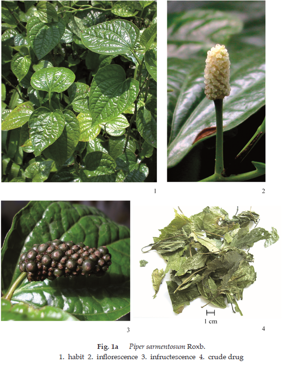
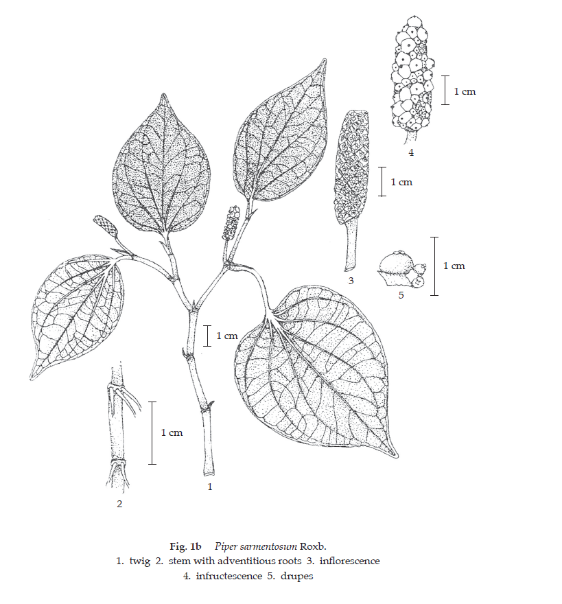
Description of the plant (Figs. 1a, 1b) Herb erect or creeping, often stoloniferous, swollen node. Leaves simple, alternate, stipulate; suborbicular ovate, or ovate-oblong, 7 to 15 cm long, 5 to 10 cm wide, surface glabrous or short hairs, apex acute to shortly acuminate, base cordate to obliquely obtuse or rounded, margin entire, slightly undulate, veins palmately 5- to 7-nerved, prominent on lower surface; petiole 2 to 5 cm long. Inflorescence leaf-opposed dense spike, generally unisexual. Male spike white, 1.5 to 3 cm long; peduncle 0.5 to 1.5 cm long; stamens 2 to 3, filament very short, anther subglobose. Female spike whitish, 2 to 5 cm long; peduncle 0.5 to 1.5 cm long; stigmas 3 to 4. Fruit drupe, obovoid, dark green when ripe. Seeds small.
Description Odour, characteristic; taste, slightly pungent.
Macroscopical (Fig. 1a) A mixture of entire and broken leaves. Entire leaf, ovate or obovate-oblong, 3.5 to 13 cm long, 2.5 to 7 cm wide; apex, acuminate; base, cordate or obtuse or obliquely obtuse; upper surface green to greenish brown, lower surface green to greyish green, lighter colour; petiole brown, 1 to 6 cm long.
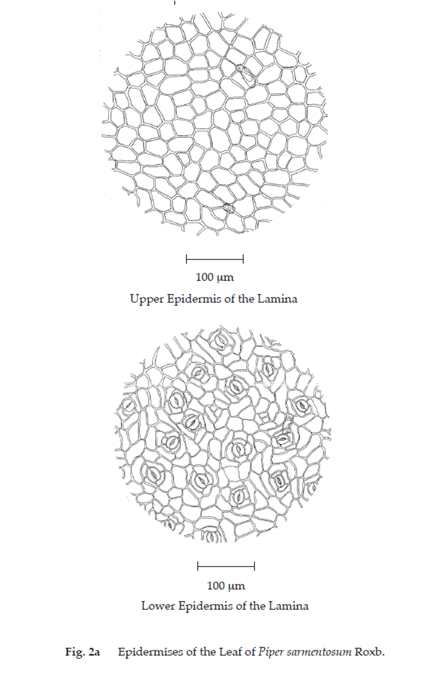
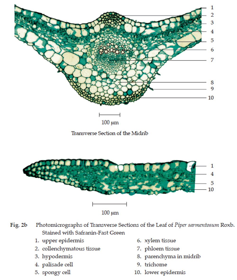
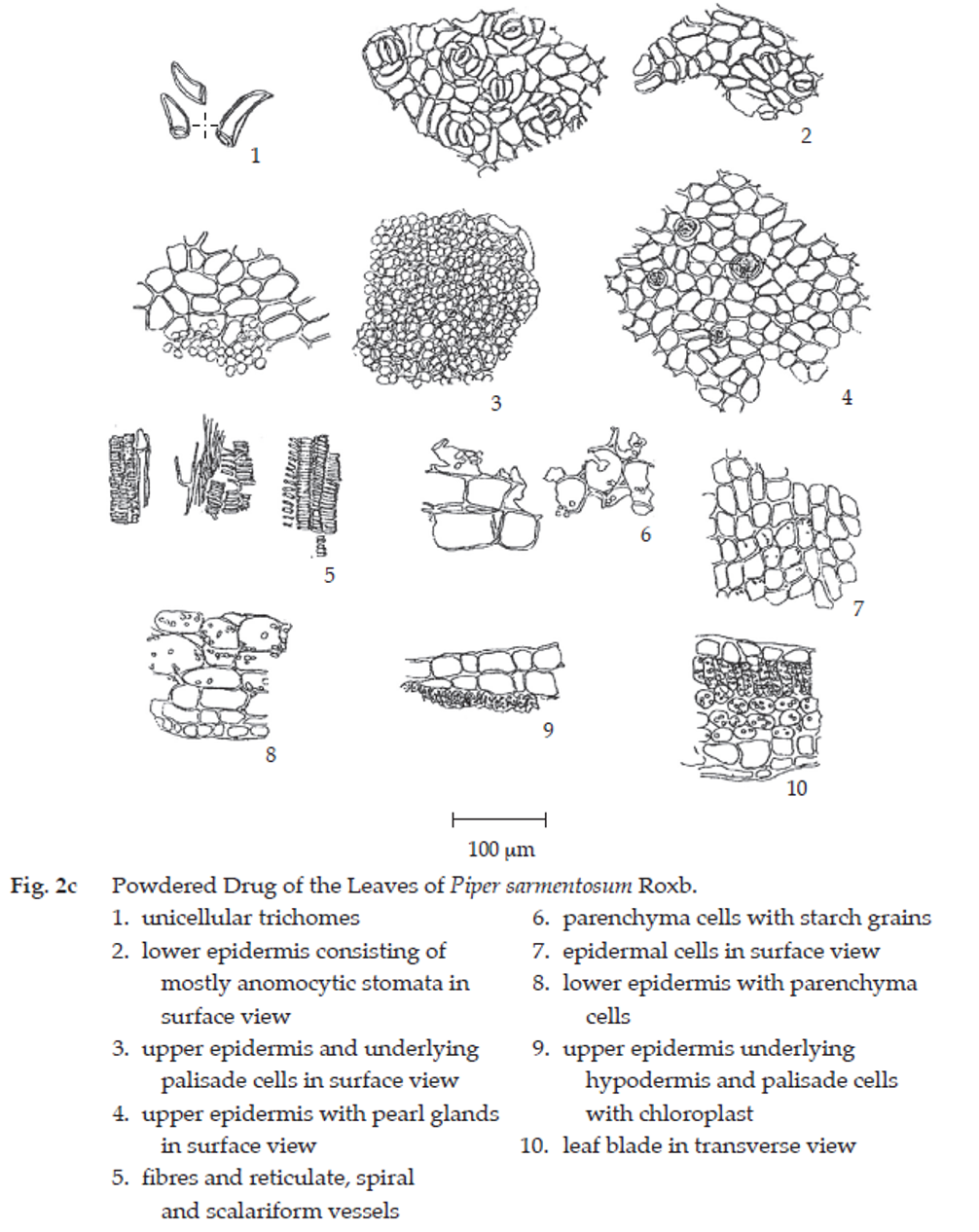
Microscopical (Figs. 2a, 2b, 2c) Transverse section of the leaf shows upper epidermis, a single layer of cuticularized rectangular cells, polygonal in surface view, with few unicellular trichomes and pearl glands. Mesophyll, a single layer of palisade parenchyma and several layers of round spongy parenchyma with scattered small vascular bundles. Lower epidermis, a single layer of rectangular cells, polygonal and irregular shape in surface view; stomata, mostly anomocytic. Hypodermis, 1 to 2 layers of cells under upper and lower epidermises of midrib and nearby area, and leaf margin.
Transverse section through the midrib of lamina shows several layers of collenchyma underneath the epidermis, parenchyma and collateral vascular bundles. Trichomes, 1 to 3 cells, uniseriate nonglandular, abundant at lower epidermis.
Piper Sarmentosum Leaf in powder possesses the diagnostic microscopical characters of the unground drug.
Packaging and storage Piper Sarmentosum Leaf shall be kept in well-closed containers, protected from light, and stored in a dry place.
Identification
A. Macerate 500 mg of the sample, in coarse powder, with 25 mL of ethanol for 24 hours, filter, and evaporate the filtrate to dryness. Dissolve the residue in 10 mL of dilute sulfuric acid and filter (solution 1). To 2 mL of solution 1, add a few drops of mercuric-potassium iodide TS: a white precipitate is produced.
B.To 2 mL of solution 1, add a few drops of modified Dragendorff TS1: an orange precipitate is produced.
C. Boil 500 mg of the sample, in coarse powder, with 10 mL of water in a water-bath for 10 minutes and filter. To 2 mL of the filtrate, add 1 or 2 drops of a 1 per cent w/v solution of iron(III) chloride: a greenish grey precipitate develops.
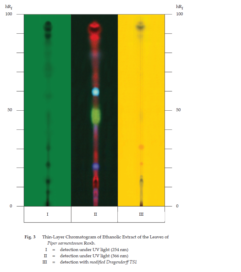
D. Carry out the test as described in the “Thin-Layer Chromatography” (Appendix 3.1), using silica gel GF254 as the coating substance and a mixture of 3 volumes of ethyl acetate and 1 volume of toluene as the mobile phase. Apply to the plate, 10 µL of the test solution prepared by macerating 500 mg of the sample, in coarse powder, with 25 mL of ethanol for 24 hours, filtering, and evaporating the filtrate to dryness. Dissolve the residue in 1 mL of ethanol. After removal of the plate, allow it to dry in air and examine under ultraviolet light (254 nm), marking the quenching spots. Subsequently examine the plate under ultraviolet light (366 nm) through the cut-off filter; several fluorescent spots of different colours are observed. Spray the plate with modified Dragendorff TS1; several spots of different colours appear (Table 1); see also Fig. 3.
| Spot | hRf Value | Detection | ||
| UV 254 | UV 366 | Modified Dragendorff TS1 | ||
| 1 | 7-9 | quenching | red | - |
| 2 | 12-14 | quenching | intense red | greyish blue |
| 3 | 14-16 | quenching | red | greyish blue |
| 4 | 21-23 | quenching | purple | orange |
| 5 | 25-27 | - | red | - |
| 6 | 30-32 | quenching | - | orange |
| 7 | 33-35 | weak quenching | - | - |
| 8 | 37-39 | - | red | - |
| 9 | 41-43 | - | light purple | - |
| 10 | 46-48 | weak quenching | green | - |
| 11 | 57-59 | weak quenching | - | - |
| 12 | 60-62 | - | intense blue | - |
| 13 | 68-70 | weak quenching | red | - |
| 14 | 71-73 | weak quenching | - | - |
| 15 | 78-80 | weak quenching | red | - |
| 16 | 88-90 | quenching | red | - |
| 17 | 92-94 | quenching | red | greyish blue |
| 18 | 95-97 | quenching | red | - |
Loss on drying Not more than 10.0 per cent w/w after drying at 105° to cons tant weight (Appendix 4.15).
Acid-insoluble ash Not more than 7.0 per cent w/w (Appendix 7.6).
Total ash Not more than 20.0 per cent w/w (Appendix 7.7).
Ethanol-soluble extractive Not less than 7.0 per cent w/w (Appendix 7.12).
Water-soluble extractive Not less than 20.0 per cent w/w (Appendix 7.12).